. 2013 Jun 28;340(6140):1236086.
doi: 10.1126/science.1236086.

Crystal structures of EF-G-ribosome complexes trapped in intermediate states of translocation

Affiliations

Crystal structures of EF-G-ribosome complexes trapped in intermediate states of translocation

Jie Zhou et al. Science. .

Abstract

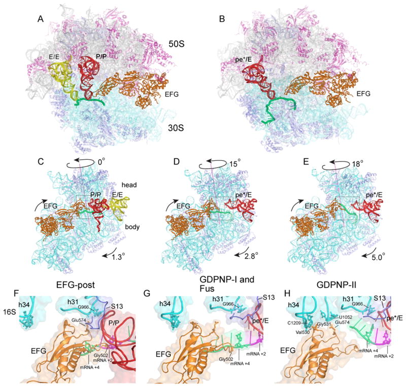
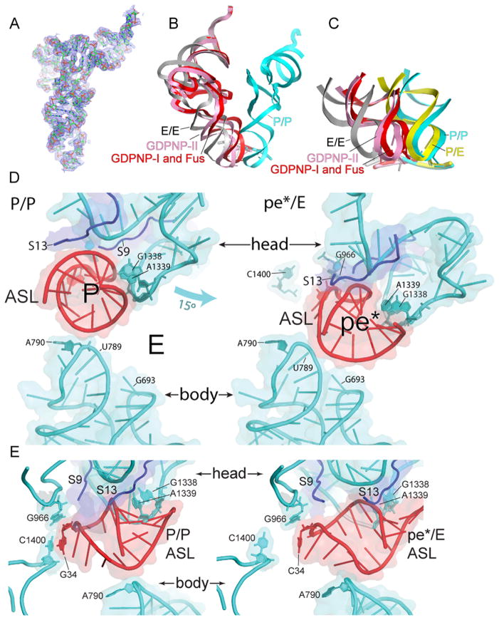
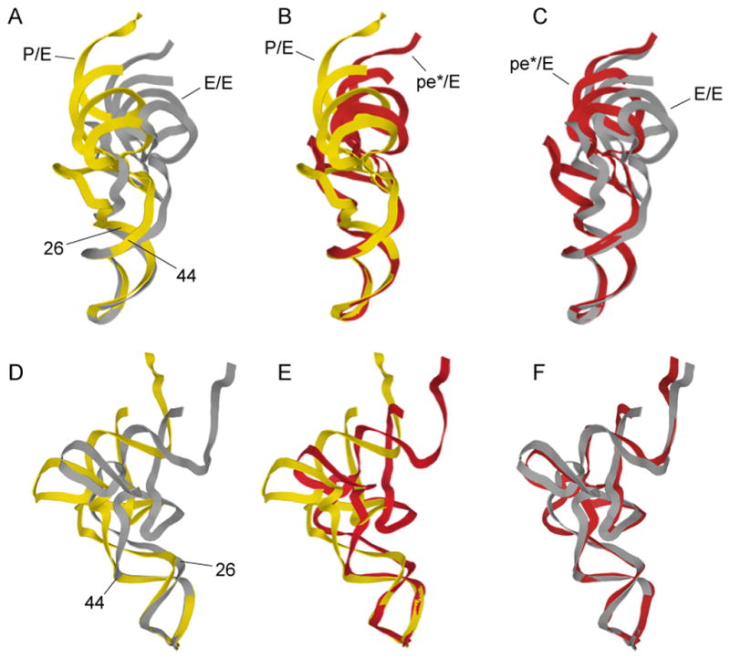
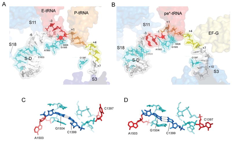
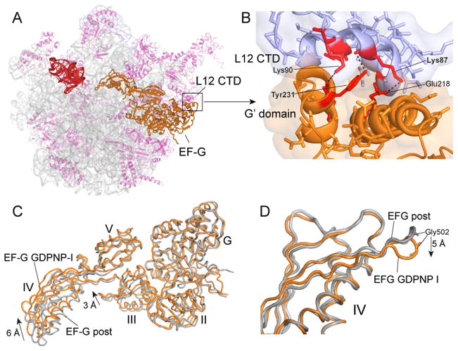
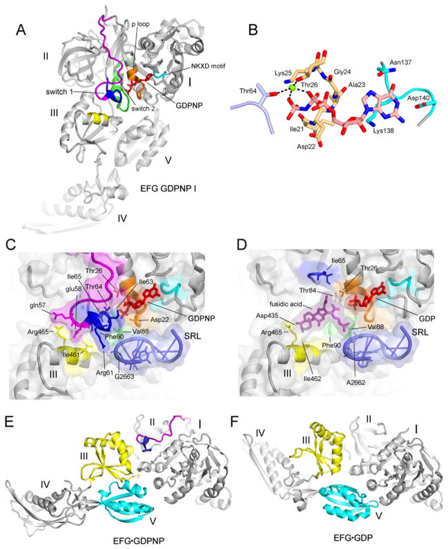
Translocation of messenger and transfer RNA (mRNA and tRNA) through the ribosome is a crucial step in protein synthesis, whose mechanism is not yet understood. The crystal structures of three Thermus ribosome-tRNA-mRNA-EF-G complexes trapped with β,γ-imidoguanosine 5'-triphosphate (GDPNP) or fusidic acid reveal conformational changes occurring during intermediate states of translocation, including large-scale rotation of the 30S subunit head and body. In all complexes, the tRNA acceptor ends occupy the 50S subunit E site, while their anticodon stem loops move with the head of the 30S subunit to positions between the P and E sites, forming chimeric intermediate states. Two universally conserved bases of 16S ribosomal RNA that intercalate between bases of the mRNA may act as "pawls" of a translocational ratchet. These findings provide new insights into the molecular mechanism of ribosomal translocation.


Figures

Figure 1
Figure 1. Structures of trapped 70S ribosome·EF-G complexes
(A,B) Overall views of the (A) non-rotated 70S·EF-G-post complex (15) and (B) Fus 70S·EF-G complex with tRNA bound in the pe*/E state. (C–E) Interface views showing 30S subunit body and head rotation in the (C) EF-G-post state (15); (D) GDPNP-I and Fus complex; and (E) GDPNP-II complex. (F–H) Close-up views of EF-G domain IV interactions with the 30S subunit head in the (F) EF-G-post complex (15); (G) GDPNP-I and Fus complex; and (H) GDPNP-II complex. 16S rRNA, cyan; 30S proteins, blue; 23S rRNA, grey; 50S proteins, magenta; mRNA, green; P/P tRNA, red; E/E tRNA, yellow; pe*/E tRNA, red; EF-G, orange.
Fig. 2
Fig. 2. Movement of tRNA from the P/P to the pe*/E state
(A) Electron density map (2Fo-Fc) contoured at 1.5σ for pe*/E tRNA from the Fus complex. (B) Superimposition of tRNA positions for the P/P (blue), E/E (grey) and pe*/E states from the Fus, (red); and GDPNP-II (pink) complexes, aligned on the 23S rRNAs from each complex. The pe*/E tRNAs move from the P site to positions midway between the P and E sites. (C) Positions of the ASLs of tRNA in the Fus (red) and GDPNP-II (pink) pe*/E states compared with those of the classical P/P (blue) and E/E (grey) (17) and hybrid-state P/E (yellow)(6) tRNAs, aligned on the 30S subunit body. (D) Interactions of (left) a P/P classical-state ASL (17) and (right) the pe*/E ASL from the 70S·EF-G Fus complex with the 30S subunit. The 15° rotation of the 30S subunit head in the EF-G opens the constriction blocking passage of the tRNA from the P site to the E site from 14Å to 22 Å, allowing translocation. (E) Side views of the complexes shown in (D). Rotation of the head moves the tRNA ASL away from its contacts with C1400 and A790 in the body and platform as it translocates into the pe*/E state.
Fig. 3
Fig. 3. Flexing of tRNA during translocation
Comparison of the conformations of tRNA in the (A,D) hybrid P/E and classical E/E states; (B,E) chimeric pe*/E and P/E states; (C,F) pe*/E and E/E states, aligned on their respective ASLs. The main site of flexing is localized to the non-canonical base pair at positions 26–42, between the D and anticodon stems. The P/E and E/E structures are from (6, 15).
Fig. 4
Fig. 4. Interactions of mRNA with the 30S subunit
(A) mRNA bound to a classical-state 70S ribosome (17). (B) mRNA bound to the Fus complex with EF-G and pe*/E tRNA. The positions of proteins S3, S11 and S18 are shown as blue transparent molecular surfaces; also shown are the positions of EF-G; the anticodon stem-loops of EF-G, P-tRNA, E-tRNA and pe*/E tRNA; the Shine-Dalgarno helix (S/D). Elements of 16S rRNA are shown in cyan. The A-, P- and E-site codons for the mRNAs during complex formation are shown in yellow, orange and red, respectively. The mRNAs are numbered with +1 corresponding to the 5′ nucleotide of the P-site codon. (C,D) The conformations of the tertiary hairpin-like structures containing the intercalating bases C1397 and A1503 (shown in red) in (C) the classical-state ribosome and (D) the 70S·EF-G Fus complex. The structures of these features in the GDPNP I and II complexes are similar to those of the Fus complex. The universally conserved C1399-G1504 base pair is shown in dark blue.
Fig. 5
Fig. 5. EF-G interactions and dynamics
(A) Overall view of the position of EF-G on the 50S subunit. (B) Detailed view of the contact surface between the G′-domain of EF-G and the C-terminal domain (CTD) of one of the four copies of protein L12. 23S rRNA (grey), 50S proteins (magenta), EF-G (orange), L12 CTD (magenta in A, blue in B). (C) Superimposition of EF-G from the fusidic acid post complex (grey) (15) with EF-G from the GDPNP-I complex (orange) by alignment on their domains I shows rearrangements in the orientation of domains III and IV. (D) Alignment of the same two EF-G structures in panel C on their respective domains IV shows local rearrangement of the loop containing the conserved Gly502-Gly503 residues (cf. Fig. 1F–H).
Fig. 6
Fig. 6. Structuring of switch loop I
(A) EF-G GDPNP-I showing the path of switch loop I (residues 40–67) which was disordered in all previous EF-G structures. The conserved core (residues 59–67) is shown in blue, and the rest of the switch loop I (residues 40–58) in magenta. (B) Structure of the GDPNP binding pocket, showing interactions with switch loop I (light blue), the guanosine recognition motif (cyan) and P loop (orange). A magnesium ion coordinating the β and γ phosphates of GDPNP is shown as a green sphere. (C) The switch I region in the GDPNP-I complex and (D) The fusidic acid binding site in the Fus complex, in the same view as for (C). In (C) and (D), showing the conserved core of switch loop I (blue) and the rest of switch loop I (magenta), guanosine recognition motif (cyan), phosphate binding loop (orange) and switch loop 2 (green) and domain III contacts (yellow); the components are shown with transparent molecular surface representations. (E) EF-G from the GDPNP-I structure containing a structured switch loop I (blue, magenta) compared with (F) the structure of free EFG·GDP containing a disordered switch loop I (27) showing movement of domains III (yellow), IV and V (cyan).

Comment in

References

    1. Moazed D, Noller HF. Nature. 1989;342:142. - PubMed
    1. Dorner S, Brunelle JL, Sharma D, Green R. Nat Struct Mol Biol. 2006;13:234. - PMC - PubMed
    1. Cornish PV, Ermolenko DN, Noller HF, Ha T. Mol Cell. 2008;30:578. - PMC - PubMed
    1. Agirrezabala X, et al. Mol Cell. 2008;32:190. - PMC - PubMed
    1. Spiegel PC, Ermolenko DN, Noller HF. RNA. 2007;13:1473. - PMC - PubMed

Publication types

MeSH terms