

Can the diagnosis of NF1 be excluded clinically? A lack of pigmentary findings in families with spinal neurofibromatosis demonstrates a limitation of clinical diagnosis
- PMID: 23812910
- PMCID: PMC3756527
- DOI: 10.1136/jmedgenet-2013-101648
Can the diagnosis of NF1 be excluded clinically? A lack of pigmentary findings in families with spinal neurofibromatosis demonstrates a limitation of clinical diagnosis
Abstract
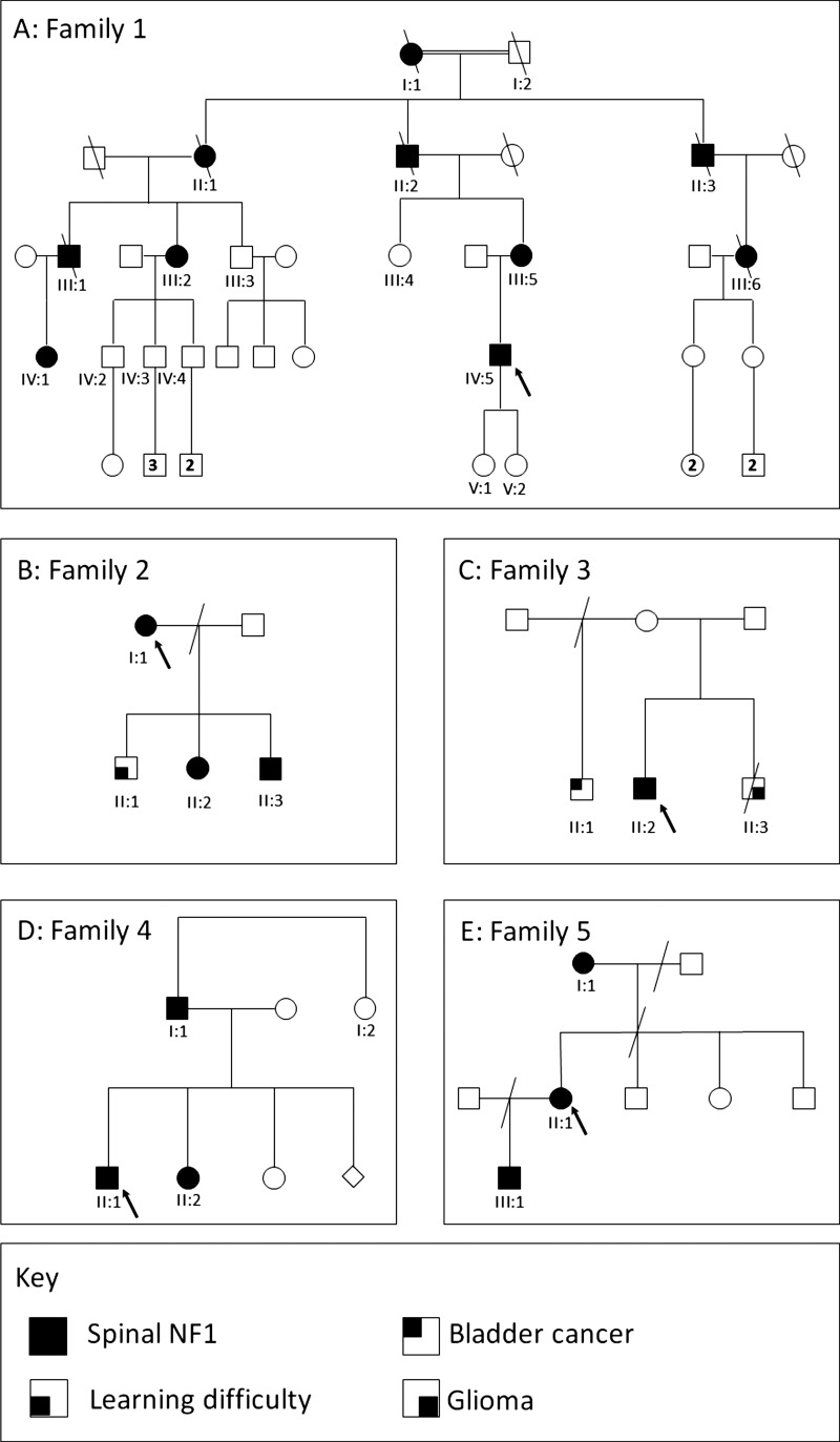
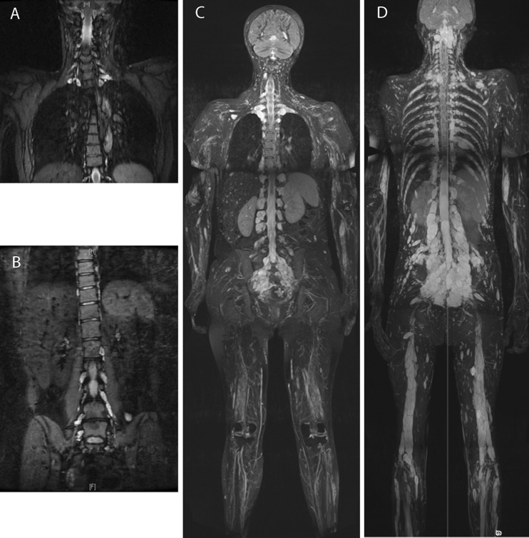
Background: Consensus clinical diagnostic criteria for neurofibromatosis type I (NF1) include café-au-lait macules and skinfold freckling. The former are frequently the earliest manifestation of NF1, and as such are of particular significance when assessing young children at risk of the condition. A phenotype of predominantly spinal neurofibromatosis has been identified in a small minority of families with NF1, often in association with a relative or absolute lack of cutaneous manifestations. An association with splicing and missense mutations has previously been reported for spinal neurofibromatosis, but on the basis of molecular results in only a few families.
Method: Patients with spinal NF1 were identified through the Manchester nationally commissioned service for complex NF1.
Results: Five families with spinal NF1 were identified, with a broad spectrum of NF1 mutations, providing further evidence that this phenotype may arise in association with any genre of mutation in this gene. Pigmentary manifestations were absent or very mild in affected individuals. Several further affected individuals, some with extensive spinal root tumours, were ascertained when additional family members were assessed.
Conclusions: Clinical NF1 consensus criteria cannot be used to exclude the diagnosis of spinal NF1, especially in childhood. This emphasises the importance of molecular confirmation in individuals and families with atypical presentations of NF1.
Keywords: Clinical genetics; Dermatology; Genetics; Other neurology.
Figures
References
-
- Viskochil D, Buchberg AM, Xu G, Cawthon RM, Stevens J, Wolff RK, Culver M, Carey J, Copeland NG, Jenkins NA, White R, O'Connell P. Deletions and a translocation interrupt a cloned gene at the neurofibromatosis type 1 locus. Cell 1990;62:187–92 - PubMed
-
- Sabol Z, Resic B, Juraski R Gjergja, Sabol F, Sizgoric M Kovac, Orsolic K, Ozretic D, Sepic-Grahovac D. Clinical sensitivity and specificity of multiple T2-hyperintensities on brain magnetic resonance imaging in diagnosis of neurofibromatosis type 1 in children: diagnostic accuracy study. Croat Med J 2011;52:488–96 - PMC - PubMed
Publication types
MeSH terms
Grants and funding
LinkOut - more resources
Full Text Sources
Other Literature Sources
Medical
Research Materials
Miscellaneous