. 2013 Jul;24(8):1250-61.
doi: 10.1681/ASN.2012121216. Epub 2013 Jul 11.

The mitochondrial-targeted compound SS-31 re-energizes ischemic mitochondria by interacting with cardiolipin

Affiliations

The mitochondrial-targeted compound SS-31 re-energizes ischemic mitochondria by interacting with cardiolipin

Alexander V Birk et al. J Am Soc Nephrol. 2013 Jul.

Abstract

Ischemia causes AKI as a result of ATP depletion, and rapid recovery of ATP on reperfusion is important to minimize tissue damage. ATP recovery is often delayed, however, because ischemia destroys the mitochondrial cristae membranes required for mitochondrial ATP synthesis. The mitochondria-targeted compound SS-31 accelerates ATP recovery after ischemia and reduces AKI, but its mechanism of action remains unclear. Here, we used a polarity-sensitive fluorescent analog of SS-31 to demonstrate that SS-31 binds with high affinity to cardiolipin, an anionic phospholipid expressed on the inner mitochondrial membrane that is required for cristae formation. In addition, the SS-31/cardiolipin complex inhibited cytochrome c peroxidase activity, which catalyzes cardiolipin peroxidation and results in mitochondrial damage during ischemia, by protecting its heme iron. Pretreatment of rats with SS-31 protected cristae membranes during renal ischemia and prevented mitochondrial swelling. Prompt recovery of ATP on reperfusion led to rapid repair of ATP-dependent processes, such as restoration of the actin cytoskeleton and cell polarity. Rapid recovery of ATP also inhibited apoptosis, protected tubular barrier function, and mitigated renal dysfunction. In conclusion, SS-31, which is currently in clinical trials for ischemia-reperfusion injury, protects mitochondrial cristae by interacting with cardiolipin on the inner mitochondrial membrane.


Figures

Figure 1.
Figure 1.
SS-31 interacts selectively with CL. (A) Chemical structures of SS-31 and [ald]SS-31. (B) Representative fluorescence emission spectra of [ald]SS-31 (1 μM, λex=360 nm) in the presence of different anionic phospholipids (3 μM). A shift in emission maximum (λmax) and increase in fluorescence intensity were observed with the addition of CL, PS, and PG but not with PA. (C) Representative fluorescence emission spectra of [ald]SS-31 in the presence of other lipids (3 μM). Chol, cholesterol. There was no shift in λmax with the zwitterionic phospholipids or cholesterol. (D) Intracellular localization of biotinylated SS-31 in feline kidney cells. Biotin was visualized with streptavidin-AlexaFluor594. (E) The interaction of CL with 1 μM [ald]SS-31 is saturable, with KD=1.87±0.64 μM. (F) Representative fluorescence emission spectra of [ald]SS-31 (1 μM) showing a concentration-dependent increase in fluorescence intensity with the addition of CL. (G) SS-31 displaces nonyl acridine orange (NAO) interaction with CL in a competitive manner. (H) Representative fluorescence emission spectra of 10 μM SS-31 (λex=254 nm) with the addition of CL at different CL:SS-31 ratios. RFU, relative fluorescence units.
Figure 2.
Figure 2.
SS-31 inhibits cyt c peroxidase activity. (A) Effects of various phospholipids on inducing cyt c peroxidase activity in vitro. PC, PE, PG, PS, or CL (30 μM) was added to 2 μM cyt c. Peroxidase activity was measured using amplex red fluorescence in the presence of 10 μM H2O2. (B) SS-31 dose-dependently inhibits cyt c peroxidase activity induced by 30 μM CL (EC50=3.5±0.03 μM). (C) Effects of various phospholipids in inducing peroxidase activity in permeabilized mitochondria. (D) SS-31 dose-dependently inhibits CL-induced peroxidase activity in permeabilized mitochondria (EC50=0.8±0.06 μM). The open square represents inhibition of peroxidase activity produced by 10 μM [ald]SS-31, showing that it has comparable activity with the activity of SS-31. (E) Effects of Ca2+ on CL-induced cyt c peroxidase activity. (F) SS-31 dose-dependently inhibits Ca2+-induced cyt c peroxidase activity (EC50=0.8±0.06 μM). All data are represented as mean ± SEM (n=4–8).
Figure 3.
Figure 3.
SS-31 prevents CL from exposing the heme Fe in cyt c to induce peroxidase activity. (A) Change in emission spectrum of [ald]SS-31 on addition of CL and cyt c. Addition of 6 μM cyt c to [ald]SS-02 (1 μM) and CL (3 μM) causes dramatic quenching and additional shift of λmax from 510 to 450 nm, suggesting that the CL–peptide complex resides in a hydrophobic domain in close proximity to the heme. (B) SS-31 prevents the effect of CL on the negative Cotton peak in Soret spectrum of cyt c. CD was carried out with 10 μM cyt c alone (gray) or in the presence of 20 μM CL (black) or CL plus 10 μM SS-31 (red).
Figure 4.
Figure 4.
SS-31 protects mitochondrial cristae during ischemia. Representative electron microscopic images of mitochondria in proximal tubular cell from sham-operated controls and saline- or SS-31–treated kidneys (A) immediately after 30 minutes of ischemia and (B) after 5 minutes of reperfusion. Original magnification, ×20,000 in top panel. The boxed area in the top panel is magnified in the corresponding middle panel. Changes in mitochondrial size and matrix density were quantified by image analysis as described in Concise Methods. Data are presented as mean ± SEM. ***P<0.001.
Figure 5.
Figure 5.
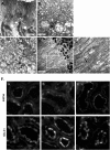
SS-31 promotes restoration of brush border in proximal tubular cells after reperfusion. Representative electron microscopic images of proximal tubules brush border from (A) sham-operated control, (B) saline-treated animal after 30 minutes of ischemia, (C) saline-treated animal after 30 minutes of ischemia and 5 minutes of reperfusion, and (D) SS-31–treated animal after 30 minutes of ischemia and 5 minutes of reperfusion. (E) Actin filaments (shown by arrows) are seen here in higher magnification from the area selected in D. (F) F-actin staining in proximal tubules of (top panel) saline and (bottom panel) SS-31 kidneys (a) at the end of 30 minutes of ischemia and after (b) 5 minutes of reperfusion and (c) 20 minutes of reperfusion. Original magnification, ×600.
Figure 6.
Figure 6.
SS-31 promotes rapid recovery of actin cytoskeleton and proximal tubular cell polarity after ischemia. Immunohistochemical staining of E-cadherin and β1-integrin in proximal tubular cells (A) in sham-operated controls, (B) after 30 minutes of ischemia, and after 5 minutes of reperfusion with (C) saline or (D) SS-31 treatment. Original magnification, ×600.
Figure 7.
Figure 7.
SS-31 prevents tubular epithelial cell detachment caused by 30 minutes of ischemia. Representative periodic acid–Schiff stain showing detachment of viable proximal tubular cells 24 hours after ischemia in saline-treated and SS-31–treated kidneys. Original magnification, ×200. Insets show higher magnifications of areas indicated by the arrows.
Figure 8.
Figure 8.
SS-31 prevents tubular cell apoptosis caused by 30 minutes of ischemia. Representative TUNEL staining in the outer medulla 24 hours after ischemia injury from (A) sham, (B) saline-treated, and (C) SS-31–treated kidneys. Original magnification, ×200. TUNEL-positive cells are indicated by arrows. Asterisks show necrotic cells. Apoptotic cells were seen in both proximal (P) and distal (D) tubules. Necrotic cells were seen more often in proximal tubules. SS-31 reduced apoptotic cells in both proximal and distal tubules. Data are represented as mean ± SEM. ***P<0.001.
Figure 9.
Figure 9.
(A) Proposed model illustrating electrostatic and hydrophobic interactions between (blue) [ald]SS-31 and (red) CL. (B) Proposed model showing how insertion of the acyl chain of CL disrupts the coordination of the heme Fe to Met80. SS peptides form a complex with CL, and the electron-dense aromatic rings help preserve the heme-Met80 coordination.

Comment in

Similar articles

Cited by

References

    1. Bagshaw SM, Laupland KB, Doig CJ, Mortis G, Fick GH, Mucenski M, Godinez-Luna T, Svenson LW, Rosenal T: Prognosis for long-term survival and renal recovery in critically ill patients with severe acute renal failure: A population-based study. Crit Care 9: R700–R709, 2005 - PMC - PubMed
    1. Bon D, Chatauret N, Giraud S, Thuillier R, Favreau F, Hauet T: New strategies to optimize kidney recovery and preservation in transplantation. Nat Rev Nephrol 8: 339–347, 2012 - PubMed
    1. Chapman JR, O’Connell PJ, Nankivell BJ: Chronic renal allograft dysfunction. J Am Soc Nephrol 16: 3015–3026, 2005 - PubMed
    1. Atkinson SJ, Hosford MA, Molitoris BA: Mechanism of actin polymerization in cellular ATP depletion. J Biol Chem 279: 5194–5199, 2004 - PubMed
    1. Sharfuddin AA, Molitoris BA: Pathophysiology of ischemic acute kidney injury. Nat Rev Nephrol 7: 189–200, 2011 - PubMed

Publication types

MeSH terms