. 2013 Dec;58(6):2176-87.
doi: 10.1002/hep.26600. Epub 2013 Oct 21.

A novel role of nucleostemin in maintaining the genome integrity of dividing hepatocytes during mouse liver development and regeneration

Affiliations

A novel role of nucleostemin in maintaining the genome integrity of dividing hepatocytes during mouse liver development and regeneration

Tao Lin et al. Hepatology. 2013 Dec.

Abstract

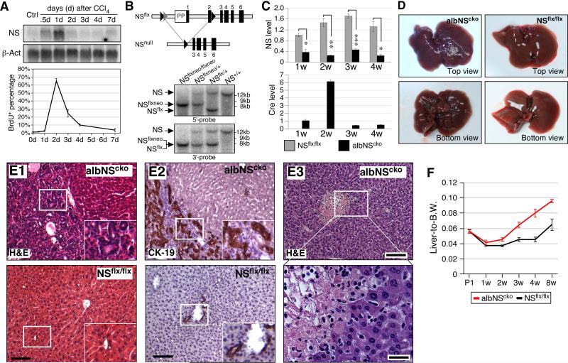
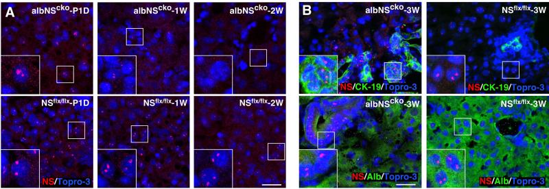
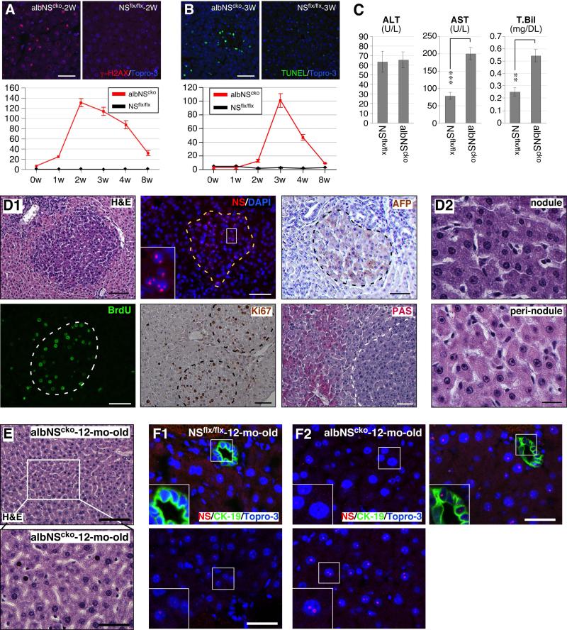
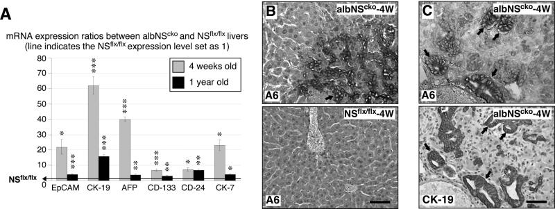
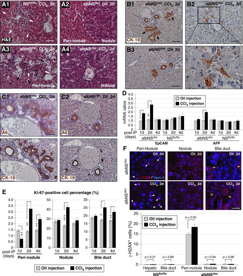
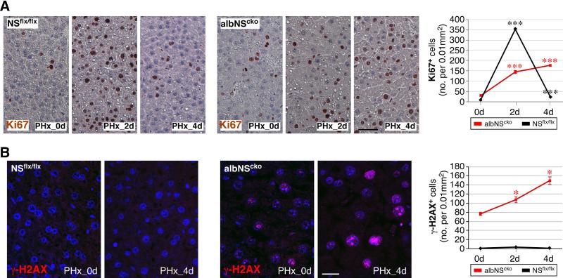
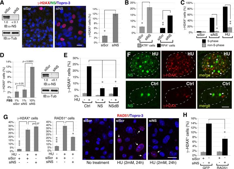
During liver development and regeneration, hepatocytes undergo rapid cell division and face an increased risk of DNA damage associated with active DNA replication. The mechanism that protects proliferating hepatocytes from replication-induced DNA damage remains unclear. Nucleostemin (NS) is known to be up-regulated during liver regeneration, and loss of NS is associated with increased DNA damage in cancer cells. To determine whether NS is involved in protecting the genome integrity of proliferating hepatocytes, we created an albumin promoter-driven NS conditional-null (albNS(cko) ) mouse model. Livers of albNS(cko) mice begin to show loss of NS in developing hepatocytes from the first postnatal week and increased DNA damage and hepatocellular injury at 1-2 weeks of age. At 3-4 weeks, albNS(cko) livers develop bile duct hyperplasia and show increased apoptotic cells, necrosis, regenerative nodules, and evidence suggestive of hepatic stem/progenitor cell activation. CCl4 treatment enhances degeneration and DNA damage in NS-deleted hepatocytes and increases biliary hyperplasia and A6(+) cells in albNS(cko) livers. After 70% partial hepatectomy, albNS(cko) livers show increased DNA damage in parallel with a blunted and prolonged regenerative response. The DNA damage in NS-depleted hepatocytes is explained by the impaired recruitment of a core DNA repair enzyme, RAD51, to replication-induced DNA damage foci.

Conclusion: This work reveals a novel genome-protective role of NS in developing and regenerating hepatocytes.


Figures

Figure 1
Figure 1. Alb-Cre-driven NS deletion causes liver damage associated with bile duct hyperplasia
(A) (Top) Northern blots show a significant upregulation of NS transcripts in CCl4-injured livers (8-week-old). The increase of NS peaks on the first day of the injection. (Bottom) the percentage of BrdU-labeled hepatocytes reaches plateau on the second day of the injection. (B) Southern blots (hybridized with 5’-probe or 3’-probe) confirm the genotypes of the NSflxneo and NSflx alleles. (C) qRT-PCR assays verify the decrease of NS (top) and the increase of Cre (bottom) in albNScko livers compared to age-matched NSflx/flx livers from 1 to 4 weeks old. (D) At 4 weeks, albNScko livers display a nodular appearance. (E1) The H&E histology of 4-week-old albNScko livers shows hepatic lobules surrounded by hyperplastic bile ducts. Insets show enlarged images of the rectangles. (E2) These hyperplastic bile ducts express CK-19. Bottom panels show images of NSflx/flx livers. (E3) From 3 weeks of age, necrotic foci can be seen in the albNScko liver parenchyma. The bottom panel shows an enlarged view of the rectangular region in the top panel. (F) The liver-to-body weight ratio of albNScko mice increases over that of NSflx/flx mice from 3 weeks old. Bar and line graphs represent mean ± SEM. *, p < 0.01, **, p < 0.001, ***, p < 0.0001. Scale bars, 100um in (E1, E2, E3 top) and 25um in (E3 bottom).
Figure 2
Figure 2. Loss of NS protein in albNScko livers by immunofluorescence
(A) A significant number of albNScko hepatocytes still retains NS expression at postnatal day 1 (P1D). Starting from 1 and 2 weeks old (1W and 2W), most albNScko hepatocytes completely lose their NS expression. (B) In 3-week-old albNScko livers (albNScko-3W), strong NS signals are found in the hyperplastic ductular epithelium (CK-19+) and absent in the Alb+ hepatocytes. In 3-week-old NSflx/flx livers (NSflx/flx-3W), NS signals are found in scattered hepatocytes but not in the bile duct epithelium. Insets show enlarged images of the square regions. Scale bars, 25um.
Figure 3
Figure 3. DNA damage is an early event of albNScko, followed by apoptosis and hepatic regeneration
(A) Alb-Cre-driven NSKO (albNScko) leads to a sharp increase of γ-H2AX+ hepatocytes that peaks at 2 weeks and declines between 3 and 8 weeks of age. Y-axis shows the number of cells per 0.01mm2 (mean ± SEM). (B) AlbNScko increases the number of apoptotic hepatocytes (green, TUNEL staining) at a later time point (3-4 weeks old). Y-axis represents cell number per 0.01mm2. (C) At 2 weeks old, albNScko mice show a significant increase in their serum levels of AST and total bilirubin (T. Bil) compared to age-matched NSflx/flx mice. The ALT levels are the same and the direct (conjugated) bilirubin levels are undetectable in both groups. ** and *** represent p values < 0.001 and 0.0001, respectively. (D1) At 2-3 weeks of age, small basophilic nodules begin to appear in the albNScko liver parenchyma (H&E). Most hepatocytes inside these nodules (demarcated by dash lines) express NS, remain mitotically active (BrdU-labeled and Ki67+), and are not fully differentiated, as indicated by their α-fetoprotein+ (AFP+) expression. The PAS staining pattern reveals loss of glycogen from the nodules. Inset shows an enlarged image of the NS signal. (D2) These newly generated hepatocytes within the nodules show basophilic cytoplasm and nuclear hyperchromasia. In contrast, the hepatocytes outside the nodules (peri-nodule) exhibit acidophilic cytoplasm, normal chromatin, and a single large nucleolus. (E) Surviving hepatocytes in 1-year-old albNScko livers display mild nuclear pleomorphism. (F) At 1 year, NSflx/flx livers show scattered NS signals in a few hepatocytes but not in CK-19+ bile duct epithelial cells (BECs) (F1), whereas albNScko livers contain regions of mostly NS-negative or low hepatocytes (F2, left upper panel) and restricted areas of strong NS-positive hepatocytes intermixed with NS-negative or low cells (F2, bottom panel). At this age, BECs of albNScko livers still show NS-positive signals (F3, right upper panel). Bars, 50um in (A, B, D1), 20um in (D2), 100um in (E top panel), and 40um in (E bottom panel, F).
Figure 4
Figure 4. AlbNScko livers show signs of hepatic stem/progenitor cell (HSPC) activation
(A) Real-time RT-PCR assays show that the transcript levels of several HSPC-related genes are increased in albNScko livers compared to their age-matched NSflx/flx controls at 4 weeks (grey) and 1 year old (black). Bar graphs show the albNScko-to-NSflx/flx ratios of HSPC-related protein expression. Asterisks represent the p values (see Fig. 1). (B) At 4 weeks old, A6+ progenitor cells (arrow) appear in the parenchyma and hyperplastic ductular areas of albNScko livers. No A6+ signal can be found in NSflx/flx livers. (C) The majority of the A6+ progenitors also coexpress the CK-19 antigen on serial sections (arrows). Scale bars, 50um.
Figure 5
Figure 5. CCl4 increases hydropic degeneration and DNA damage in NS-depleted hepatocytes and enhances A6+ cell expansion and biliary hyperplasia in albNScko livers
(A) Mice are injected with CCl4 at 2 weeks of age and analyzed on the first, second, and fourth day post injection. H&E staining shows that CCl4 causes acute pericentral necrosis and leukocyte infiltration (indicated by white arrows) in both NSflx/flx (A1) and albNScko livers (A3) during the first 2 days of the injection. CCl4 triggers hydropic degeneration (indicated by black arrows) of hepatocytes specifically in the peri-nodular regions of albNScko livers. Bars, 100um. (B) In response to CCl4 treatment, albNScko livers also display a notable increase of CK-19+ ducts (B1, arrows) and scattered progenitor-like cells (B2), both of which are not seen in CCl4-treated NSflx/flx livers (B3). (C) Immunostaining on serial sections of 4um thickness shows that A6 and CK-19 double-positive progenitor cells are increased significantly in CCl4-treated albNScko livers (C1) compared to oil-treated albNScko livers (C2). (D) qRT-PCR assays showed that CCl4-induced damage upregulates the transcript levels of both EpCAM and AFP in albNScko livers but not in NSflx/flx livers. Asterisks represent p values (described in Fig. 1). (E) In oil-treated albNScko livers (grey bars), most Ki67+ cells are found in the regenerative nodules and the bile duct epithelium, and only a small percentage of the non-regenerative hepatocytes in peri-nodular regions are Ki-67+. Following CCl4 treatment, mitotic cells are increased most significantly on the second day post injection in the peri-nodular areas, regenerative nodules, and bile duct epithelium. (F) Most γ-H2AX+ cells (indicated by white arrows) in oil-treated albNScko livers are found in the peri-nodular areas. On the second day post injection, CCl4 increases γ-H2AX+ cells only in the peri-nodular regions of albNScko livers where NS is deleted but not in the regenerative nodules or the bile duct epithelium (indicated by yellow arrows). The CCl4 treatment itself does not cause any DNA damage in NSflx/flx livers. Scale bars, 50um in (A, B, and C) and 20um in (F).
Figure 6
Figure 6. 70% partial hepatectomy (PHx) increases DNA damage in hepatocytes in 4-week-old albNScko livers
Mice received PHx at 4 weeks of age, and their liver samples were analyzed before the operation (PHx_0d), on the second day (PHx_2d) and fourth day (PHx_4d) after the operation. (A) In response to PHx, NSflx/flx livers show a significant increase of Ki67+ cells that peaks on the second day and recovers mostly on the fourth day. Before the operation, albNScko livers contain more Ki67+ cells than NSflx/flx livers. Following PHx, albNScko livers show a blunted and prolonged regenerative response compared to NSflx/flx livers. (B) Two days after PHx, albNScko livers show a significant increase of γ-H2AX+ cells and the number of γ-H2AX+ cells continues to increase 4 days after PHx (PHx_4d). In contrast, PHx does not increases γ-H2AX+ cells in NSflx/flx livers. Y-axis shows the number of γ-H2AX+ cells per 0.01mm2 (mean ± SEM). * and *** indicate p < 0.05 and 0.0001, respectively. Scale bars, 50um in (A) and 20um in (B).
Figure 7
Figure 7. NS depletion triggers replication-dependent DNA damage and impairs RAD51 recruitment to HU-induced foci in proliferating hepatocytes
(A) Compared to control knockdown by siScr, NS-knockdown (NSKD) by siNS increases the percentage of γ-H2AX+ cells (red) in primary hepatocytes isolated from 2-week-old NSflx/flx livers. Samples were co-labeled with anti-NS (green) and Topro-3 staining (blue). The knockdown efficiency of NS protein is >90%. Scale bar, 20um. (B) NSKD also increases the percentages of ATR+ (grey bars) and RPA32+ hepatocytes (black bars). (C) NSKD has a much stronger effect in triggering DNA damage (γ-H2AX+ foci) in S-phase cells (BrdU+) than in non-S-phase cells. This DNA damage profile resembles the effect of HU. (D) NSKD elicits little or no DNA damage in hepatocytes grown under the low serum (1%) condition, which lowers the proliferative rate of hepatocytes without reducing the NSKD efficiency. (E) Overexpression of wildtype NS or NSdB (a nucleoplasmic NS mutant) can both protect primary hepatocytes from HU-induced γ-H2AX+ damage foci. (F) HU treatment of primary hepatocytes induces foci formation of endogenous NS (Ab2438, green) in the nucleoplasm, some of which colocalizes with the γ-H2AX signal (red). Arrows indicate the nucleoli. Scale bars, 5um. (G) In siScr-KD hepatocytes, HU treatment significantly increases the percentages of γ-H2AX+ and RAD51+ foci. In NS-depleted hepatocytes, HU increases the percentage of γ-H2AX+ cells, but its effect on triggering RAD51 foci formation is greatly attenuated. Arrows indicate cells with RAD51+ foci. (H) Overexpression of RAD51 can partially rescue the DNA damage (γ-H2AX+) phenotype of NS-depleted hepatocytes. Bar graphs, mean ± SEM.

References

    1. Saintigny Y, Delacote F, Vares G, Petitot F, Lambert S, Averbeck D, Lopez BS. Characterization of homologous recombination induced by replication inhibition in mammalian cells. Embo J. 2001;20:3861–3870. - PMC - PubMed
    1. Petermann E, Helleday T. Pathways of mammalian replication fork restart. Nat Rev Mol Cell Biol. 2010;11:683–687. - PubMed
    1. Helleday T. Amplifying tumour-specific replication lesions by DNA repair inhibitors - a new era in targeted cancer therapy. Eur J Cancer. 2008;44:921–927. - PubMed
    1. Ciccia A, Elledge SJ. The DNA damage response: making it safe to play with knives. Mol Cell. 2010;40:179–204. - PMC - PubMed
    1. Tsai RY, McKay RD. A nucleolar mechanism controlling cell proliferation in stem cells and cancer cells. Genes Dev. 2002;16:2991–3003. - PMC - PubMed

Publication types

MeSH terms