. 2013 Sep;28(6):551-9.
doi: 10.1177/1533317513494457. Epub 2013 Jun 28.

Vascular risk factors: a ticking time bomb to Alzheimer's disease

Affiliations

Vascular risk factors: a ticking time bomb to Alzheimer's disease

Jack C de la Torre. Am J Alzheimers Dis Other Demen. 2013 Sep.

Abstract

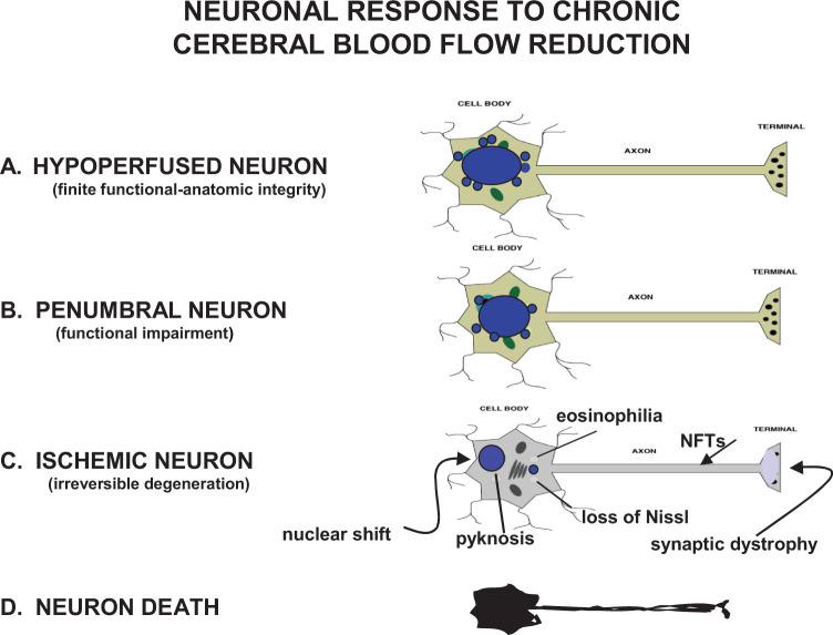
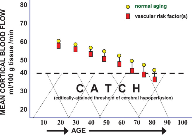
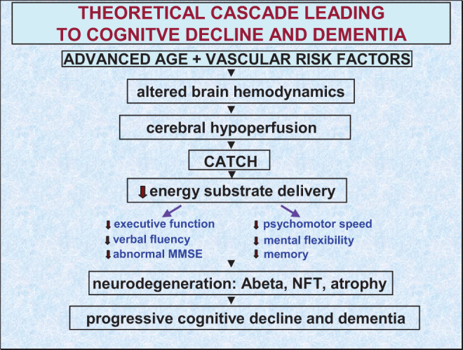
Evidence is growing that vascular risk factors (VRFs) for Alzheimer's disease (AD) affect cerebral hemodynamics to launch a cascade of cellular and molecular changes that initiate cognitive deficits and eventual progression of AD. Neuroimaging studies have reported VRFs for AD to be accurate predictors of cognitive decline and dementia. In regions that participate in higher cognitive function, middle temporal, posterior cingulate, inferior parietal and precuneus regions, and neuroimaging studies indicate an association involving VRFs, cerebral hypoperfusion, and cognitive decline in elderly individuals who develop AD. The VRF can be present in cognitively intact individuals for decades before mild cognitive deficits or neuropathological signs are manifested. In that sense, they may be "ticking time bombs" before cognitive function is demolished. Preventive intervention of modifiable VRF may delay or block progression of AD. Intervention could target cerebral blood flow (CBF), since most VRFs act to lower CBF in aging individuals by promoting cerebrovascular dysfunction.

Keywords: Alzheimer’s; aging; cerebral blood flow; hypoperfusion; neuroimaging; vascular risk factors.


Conflict of interest statement

The author declared no potential conflicts of interest with respect to the research, authorship, and/or publication of this article.

Figures

Figure 1.
Figure 1.
Four basic neuronal states generated by vascular pathology that reduces cerebral blood flow (CBF) supply and alters normal hemodynamic response to local or global ischemia. A, Hypoperfused neurons with blood flow values exceeding 22 mL/100 g/min but still below normal blood flow for gray matter; (B) penumbral neurons with blood flows ranging at 12 to 22 mL/100 g/min; (C) ischemic neurons with blood flow values under 12 mL/100 g/min. Very few, if any, structural changes are seen in hypoperfused neurons but their function may undergo progressive impairment. Penumbral neurons, also referred to as moderate ischemia, generally display mild structural changes and moderate functional deficits but are still capable of recovery assuming appropriate intervention is applied. Ischemic neurons can quickly reach the point of no return where the cell becomes unable to recover its normal morphology and function even if all processes leading to death are interventionally arrested. Acute ischemic neurons typically show cytoplasmic eosinophilia, dispersion of Nissl substance, nuclear shrinking (pyknosis), and nuclear displacement to the periphery. When ischemic neurons are chronically injured, as in neurodegeneration, no eosinophilia is seen and instead, the cell body shrinks and becomes more angular, synaptic damage can be seen, and axonal neurofibrillary tangles (NFTs) may dominate. D, Neuronal death is the state where permanent and complete arrest of energy metabolism occurs.
Figure 2.
Figure 2.
Normal resting cerebral blood flow (CBF) is approximately 65 mL/100g/min at a young age. The CBF normally declines 0.5% per year so from age 20 to 60, a 20% reduction in brain blood flow is normally expected (circles). In patients with mild cognitive impairment (MCI), CBF can further drop an additional 15% to 20%, but the cause for this CBF fall is unclear. Critically attained threshold of cerebral hypoperfusion (CATCH) argues that vascular risk factors in the presence of advanced aging can add another 15% reduction in CBF resulting in a brain metabolic energy crisis due to chronic, subnormal delivery of energy nutrients needed to sustain the high energy requirement of brain cell consumption.
Figure 3.
Figure 3.
Theoretical cascade of events leading to cognitive failure and Alzheimer or vascular dementia. The cascade may begin with a double drop in cerebral blood flow (see Figure 2) during normal aging and in the presence of vascular risk factors. These 2 events are reported to alter brain hemodynamics giving rise to chronic cerebral hypoperfusion. With time, critically attained threshold of cerebral hypoperfusion (CATCH) is reached where brain cells, especially those sensitive to ischemia in the inferior temporal region, precuneus, and posterior cingulate cortex can no longer cope with reduced energy substrate delivery. The outcome of this process affects executive function, verbal fluency, psychomotor speed, and mental flexibility, which may progress to mild cognitive impairment, severe neurodegeneration, and dementia.

Similar articles

Cited by

References

    1. Buxton R. Introduction to Functional Magnetic Resonance Imaging: Principles and Techniques. Cambridge, England: Cambridge University Press; 2002.
    1. Leenders KL, Perani D, Lammertsma AA, et al. Cerebral blood flow, blood volume and oxygen utilization. Normal values and effect of age. Brain. 2009;113(pt 1):27–47. - PubMed
    1. Pantano P, Baron JC, Lebrun-Grandie P, Duquesnoy N, Bousser MG, Comar D. Regional cerebral blood flow and oxygen consumption in human aging. Stroke. 1994;15(4):635–641. - PubMed
    1. Zou Q, Wu CW, Stein EA, Zang Y, Yang Y. Static and dynamic characteristics of cerebral blood flow during the resting state. Neuroimage. 2009;48(3):515–524. - PMC - PubMed
    1. Gur RC, Gur RE, Obrist WD, Skolnick BE, Reivich M. Age and regional cerebral blood flow at rest and during cognitive activity. Arch Gen Psychiatry. 1987;44(7):617–621. - PubMed