

Dermatophytosis in cats: ABCD guidelines on prevention and management
- PMID: 23813824
- PMCID: PMC11148949
- DOI: 10.1177/1098612X13489222
Dermatophytosis in cats: ABCD guidelines on prevention and management
Abstract
Overview: Dermatophytosis, usually caused by Microsporum canis, is the most common fungal infection in cats worldwide, and one of the most important infectious skin diseases in this species. Many adult cats are asymptomatic carriers. Severe clinical signs are seen mostly in kittens or immunosuppressed adults. Poor hygiene is a predisposing factor, and the disease may be endemic in shelters or catteries. Humans may be easily infected and develop a similar skin disease.
Infection: Infectious arthrospores produced by dermatophytes may survive in the environment for about a year. They are transmitted through contact with sick cats or healthy carriers, but also on dust particles, brushes, clothes and other fomites.
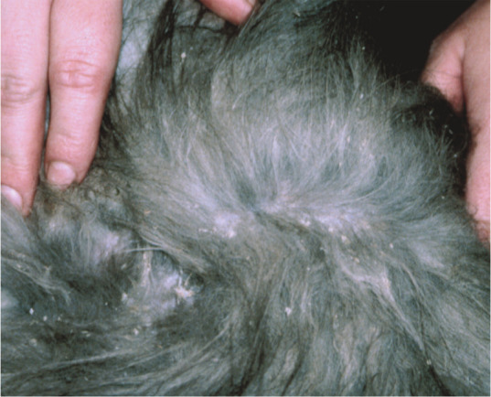
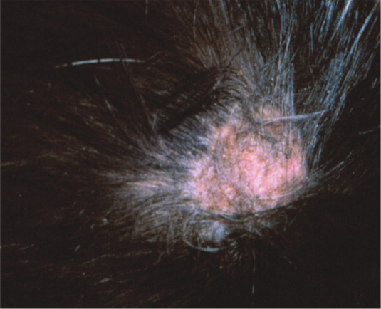
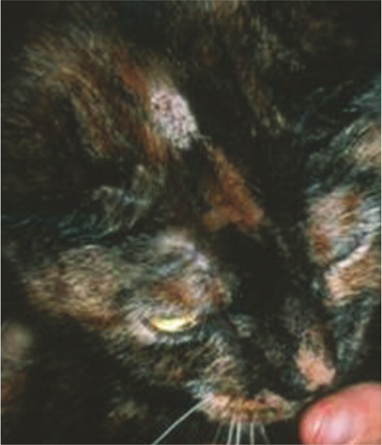
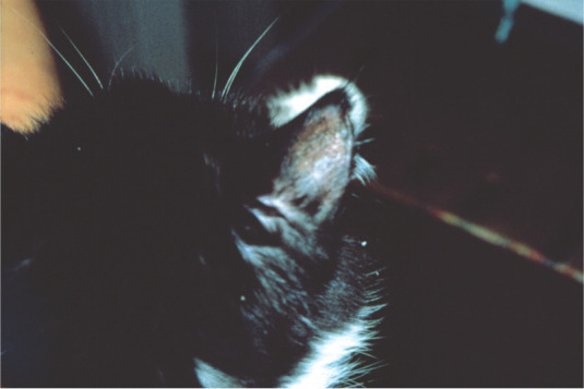
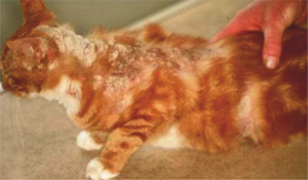
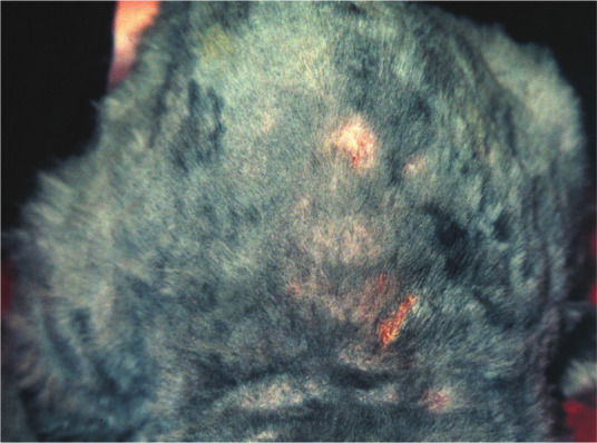
Disease signs: Circular alopecia, desquamation and sometimes an erythematous margin around central healing ('ringworm') are typical. In many cats this is a self-limiting disease with hair loss and scaling only. In immunosuppressed animals, the outcome may be a multifocal or generalised skin disease.
Diagnosis: Wood's lamp examination and microscopic detection of arthrospores on hairs are simple methods to confirm M canis infection, but their sensitivity is relatively low. The gold standard for detection is culture on Sabouraud agar of hairs and scales collected from new lesions.
Disease management: In shelters and catteries eradication is difficult. Essential is a combination of systemic and topical treatments, maintained for several weeks. For systemic therapy itraconazole is the drug of choice, terbinafine an alternative. Recommended topical treatment is repeated body rinse with an enilconazole solution or miconazole with or without chlorhexidine. In catteries/shelters medication must be accompanied by intensive decontamination of the environment.
Vaccination: Few efficacy studies on anti-M canis vaccines (prophylactic or therapeutic) for cats have been published, and a safe and efficient vaccine is not available.
Conflict of interest statement
The authors do not have any potential conflicts of interest to declare.
Figures
References
-
- Moriello KA, DeBoer DJ. Dermatophytosis. In: Greene CE. (ed). Infectious diseases of the dog and cat. 4th ed. St Louis: Elsevier, 2012, pp 588–602.
-
- Sparkes AH, Werrett G, Stokes CR, Gruffyd-Jones TJ. Microsporum canis: inapparent carriage by cats and the viability of arthrospores. J Small Anim Pract 1994; 35: 397–401.
-
- Mancianti F, Giannelli C, Bendinelli M, Poli A. Mycological findings in feline immunodeficiency virus-infected cats. J Med Vet Mycol 1992; 30: 257–259. - PubMed
-
- Sierra P, Guillot J, Jacob H, Bussiéras S, Chermette R. Fungal flora on cutaneous and mucosal surfaces of cats infected with feline immunodeficiency virus or feline leukemia virus. Am J Vet Res 2000; 61: 158–161. - PubMed
-
- Mignon BR, Losson B. Prevalence and characterization of Microsporum canis carriage in cats. J Med Vet Mycol 1997; 35: 249–256. - PubMed
Publication types
MeSH terms
Substances
Grants and funding
LinkOut - more resources
Full Text Sources
Other Literature Sources
Medical
Miscellaneous
