

Cryptococcosis in cats: ABCD guidelines on prevention and management
- PMID: 23813826
- PMCID: PMC11148960
- DOI: 10.1177/1098612X13489224
Cryptococcosis in cats: ABCD guidelines on prevention and management
Abstract
Overview: Cryptococcosis is worldwide the most common systemic fungal disease in cats; it is caused by the Cryptococcus neoformans- Cryptococcus gattii species complex, which includes eight genotypes and some subtypes (strains) with varying geographical distribution, pathogenicity and antimicrobial susceptibility. Cats acquire the infection from a contaminated environment. The prognosis is favourable in most cases, provided a diagnosis is obtained sufficiently early and prolonged treatment is maintained.
Infection: Basidiospores are the infectious propagules of Cryptococcus species as they penetrate the respiratory system and induce primary infection. Asymptomatic colonisation of the respiratory tract is more common than clinical disease. Avian guanos, particularly pigeon droppings, offer favourable conditions for the reproduction of C neoformans. Both Cryptococcus species are associated with decaying vegetation.
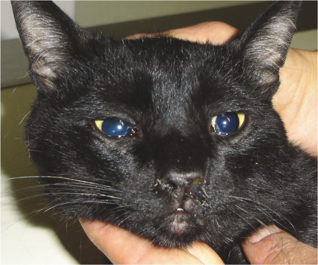
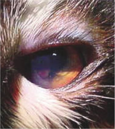
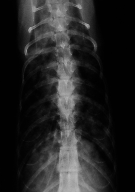
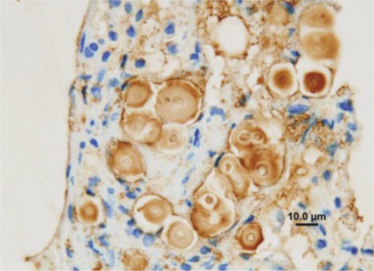
Disease signs: Cryptococcosis caused by C neoformans or C gattii is indistinguishable clinically. The disease can present in nasal, central nervous system (which can derive from the nasal form or occur independently), cutaneous and systemic forms.
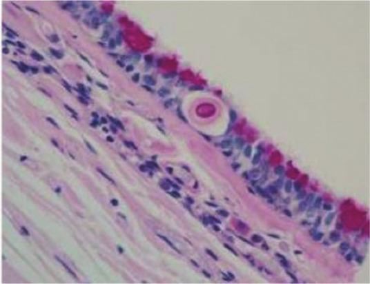
Diagnosis: An easy and reliable test for cryptococcosis diagnosis is antigen detection in body fluids. Only isolation and polymerase chain reaction allow identification of the species genotype.
Disease management: Amphotericin B, ketoconazole, fluconazole and itraconazole have all been used to treat cats. Surgical excision of any nodules in the skin, nasal or oral mucosa assists recovery. Continued treatment is recommended until the antigen test is negative.
Prevention: Efficient preventive measures have not been demonstrated. Vaccines are not available.
Conflict of interest statement
The authors do not have any potential conflicts of interest to declare.
Figures


References
-
- Sykes JE, Malik R. Cryptococcosis. In: Greene CE. (ed). Infectious diseases of the dog and cat. 4th ed. St Louis: Saunders, Elsevier, 2012, pp 621–634.
-
- Lester SJ, Malik R, Bartlett KH, Duncan CG. Cryptococcosis: update and emergence of Cryptococcus gattii. Vet Clin Pathol 2011; 40: 4–17. - PubMed
-
- Alspaugh JA, Davidson RC, Heitman J. Morphogenesis of Cryptococcus neoformans. Contrib Microbiol 2000; 5: 217–238. - PubMed
-
- Lin X, Heitman J. The biology of the Cryptococcus neoformans species complex. Annu Rev Microbiol 2006; 60: 69–105. - PubMed
-
- Kano R, Kitagawat M, Oota S, Oosumit T, Murakami Y, Tokuriki M, et al.. First case of feline systemic Cryptococcus albidus infection. Med Mycol 2008; 46: 75–77. - PubMed
Publication types
MeSH terms
Substances
Grants and funding
LinkOut - more resources
Full Text Sources
Other Literature Sources
Miscellaneous
