. 2014 Mar;59(3):898-910.
doi: 10.1002/hep.26592. Epub 2014 Jan 30.

NLRP3 inflammasome activation results in hepatocyte pyroptosis, liver inflammation, and fibrosis in mice

Affiliations

NLRP3 inflammasome activation results in hepatocyte pyroptosis, liver inflammation, and fibrosis in mice

Alexander Wree et al. Hepatology. 2014 Mar.

Abstract

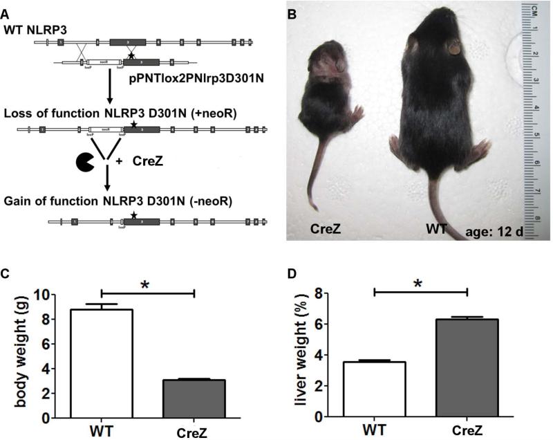
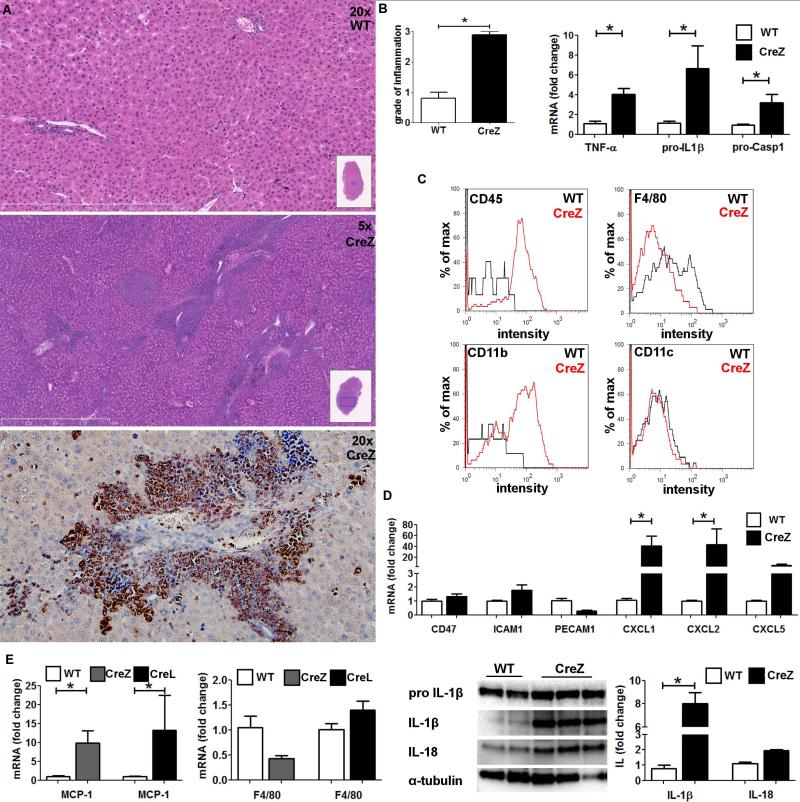
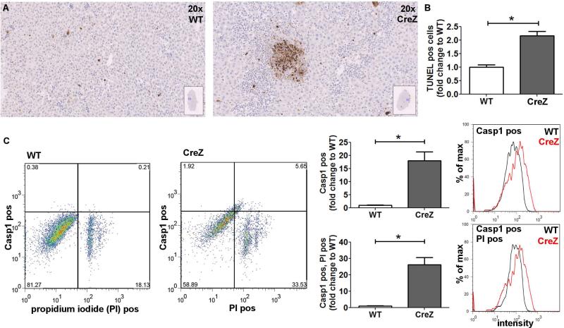
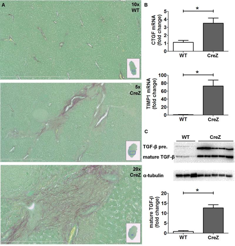
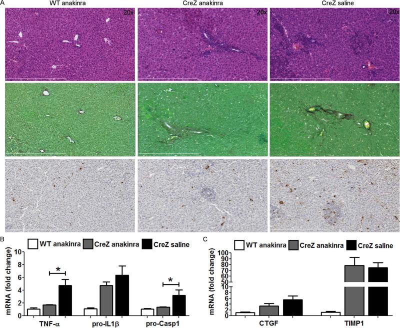
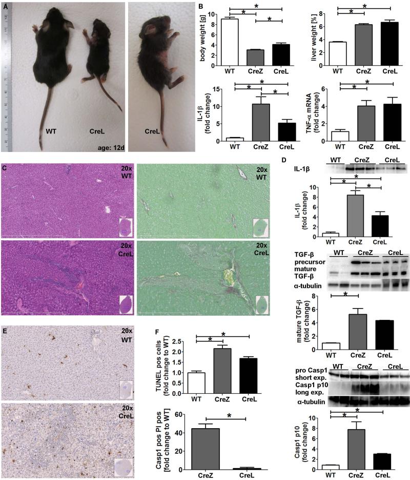
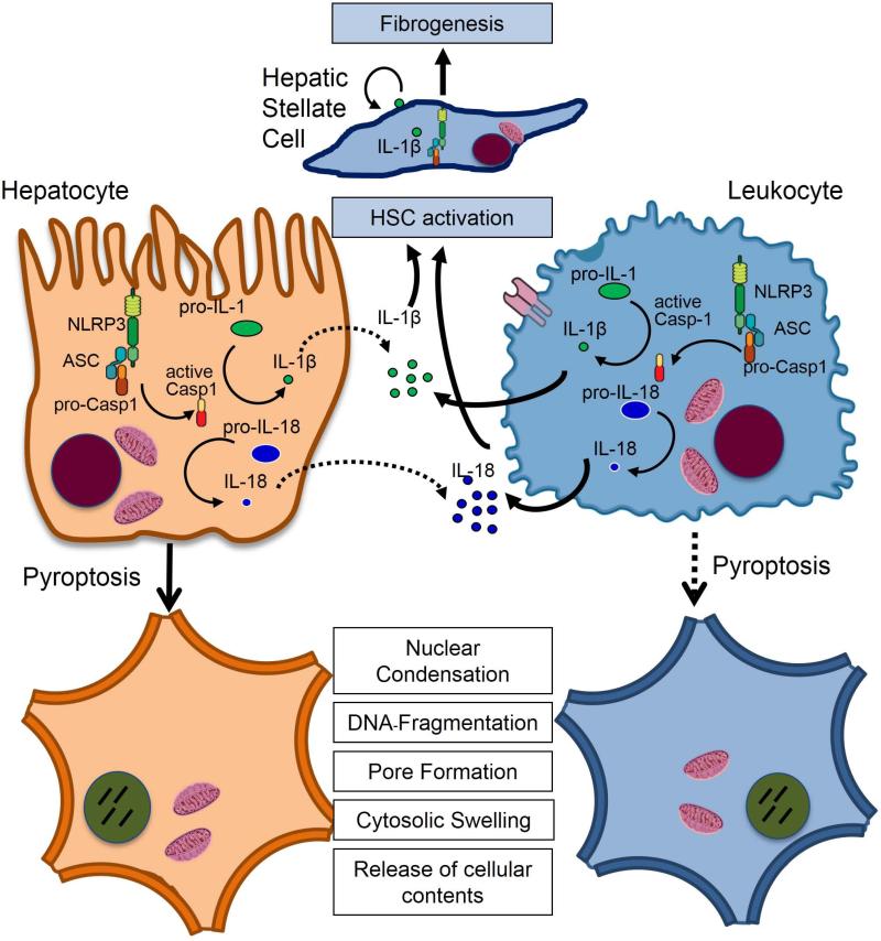
Inflammasome activation plays a central role in the development of drug-induced and obesity-associated liver disease. However, the sources and mechanisms of inflammasome-mediated liver damage remain poorly understood. Our aim was to investigate the effect of NLRP3 inflammasome activation on the liver using novel mouse models. We generated global and myeloid cell-specific conditional mutant Nlrp3 knock-in mice expressing the D301N Nlrp3 mutation (ortholog of D303N in human NLRP3), resulting in a hyperactive NLRP3. To study the presence and significance of NLRP3-initiated pyroptotic cell death, we separated hepatocytes from nonparenchymal cells and developed a novel flow-cytometry-based (fluorescence-activated cell sorting; FACS) strategy to detect and quantify pyroptosis in vivo based on detection of active caspase 1 (Casp1)- and propidium iodide (PI)-positive cells. Liver inflammation was quantified histologically by FACS and gene expression analysis. Liver fibrosis was assessed by Sirius Red staining and quantitative polymerase chain reaction for markers of hepatic stellate cell (HSC) activation. NLRP3 activation resulted in shortened survival, poor growth, and severe liver inflammation; characterized by neutrophilic infiltration and HSC activation with collagen deposition in the liver. These changes were partially attenuated by treatment with anakinra, an interleukin-1 receptor antagonist. Notably, hepatocytes from global Nlrp3-mutant mice showed marked hepatocyte pyroptotic cell death, with more than a 5-fold increase in active Casp1/PI double-positive cells. Myeloid cell-restricted mutant NLRP3 activation resulted in a less-severe liver phenotype in the absence of detectable pyroptotic hepatocyte cell death.

Conclusions: Our data demonstrate that global and, to a lesser extent, myeloid-specific NLRP3 inflammasome activation results in severe liver inflammation and fibrosis while identifying hepatocyte pyroptotic cell death as a novel mechanism of NLRP3-mediated liver damage.


Figures

Fig. 1
Fig. 1. Nlrp3 knock-ins show growth impairment
Nlrp3 CreZ knock-in pups - with the aspartate 301 to asparagine (D301N) substitution (A) – were often indistinguishable from wild type (WT) siblings at birth but showed growth retardation with significantly lower body weight after 2 weeks (B,C). Notably, Nlrp3 mutants showed significant higher liver weight as a percentage of body weight than WTs; Nlrp3 CreZ median 6.3%, WT 3.5% (D); (n≥7 for each group).
Fig. 2
Fig. 2. Global mutant NLPR3 leads to severe liver inflammation
Liver histology of global Nlrp3 knock-ins (CreZ) revealed severe inflammation in comparison to WT littermates, with infiltrating cells being mainly myeloperoxidase positive and therefore classified as neutrophils (representative of 7-13 in each group) (A). Conclusively liver samples of Nlrp3 CreZ mice exhibited significantly higher grade of inflammation scores (B). Moreover, analysis of mRNA levels within whole liver samples showed significantly higher yields of pro-IL1β (Nlrp3 CreZ 6.6-fold change to WT; p<0.05), pro-Casp1 (3.2-fold; p<0.05), and TNF-α (4.0-fold; p<0.05) than WTs (n≥7 for each group) (B). After cell separation infiltrating cells (red curve) of Nlrp3 CreZ mutants showed high intensity in CD45 and CD11b staining compared to WT mice (gray curve); no difference was observed in CD11c, CD4, and CD8 staining; and Nlrp3 CreZ mutants showed less F4/80 intensity (n≥7 for each group) (C). The neutrophil leukocyte recruitment was mediated via a marked increase in chemoattractants as CXCL1, CXCL2 and CXCL5 whereas endothelium expressed leukocyte adhesion molecules (CD47, ICAM1, PECAM1) did not show significant alterations in Nlrp3 CreZ mice (D) (n≥4 for each group). Analysis of whole liver samples showed no significant differences in F4/80 mRNA levels even though the monocyte chemoattractant protein 1 (MCP-1) was significantly higher in mutant mice with global (CreZ) and myeloid specific (CreL) inflammasome activation (n≥7 for each group) (E). IL-1β and IL-18 protein levels were markedly elevated in whole liver lysates of Nlrp3 CreZ mice (n≥3 for each group) (E).
Fig. 3
Fig. 3. Global NLRP3 inflammasome activation results in hepatocyte pyroptosis
TUNEL staining of Nlrp3 CreZ mutant liver samples showed significantly more positive cells than WTs. Livers from Nlrp3 CreZ mice displayed TUNEL positive cells throughout the tissue with several dense areas of TUNEL positive cells indicative of pyroptotic cell death (n≥5 for each group) (A, B). Conclusively, Nlrp3 CreZ mutant mice showed more Casp1 and PI double-positive hepatocytes (upper right quadrant) than WT - representative scatterplots are shown in panel C. An approximately fifteenfold increase in Casp1 positive cells was detected in Nlrp3 CreZ (red curve) in comparison to WT (gray curve), the entire population of hepatocytes was analyzed. Casp1 and PI double-positive cells were also detected at a twentyfold increased level in hepatocytes of Nlrp3 CreZ mutants; (n≥7 for each group).
Fig. 4
Fig. 4. NLRP3 increases hepatic stellate cell (HSC) activation and spontaneous collagen deposition
Nlrp3 mutants showed increased collagen deposition in comparison to WT littermates visualized by Sirius red staining in close proximity to inflammatory hot spots (A). Moreover, Nlrp3 mutants showed significantly higher CTGF and TIMP1 mRNA levels compared to WT mice (B). TGF-β precursor protein as well as mature TGF-β also showed significantly higher values in Nlrp3 CreZ mice in comparison to WT mice (C); (n≥7 for each group)
Fig. 5
Fig. 5. Treatment with IL-1 receptor antagonist partially attenuates liver pathology in Nlrp3 CreZ mice
Nlrp3 CreZ mutant mice and WT were treated with 300 mg/kg IL-1 receptor antagonist (anakinra) or saline from day 2 to 13. Liver histology of anakinra treated mutants showed a marked decrease in liver inflammation and TUNEL positive cells, whereas collagen deposition assessed by sirius red staining was unaffected (A). Correspondingly, mRNA levels of TNF-α and pro-Casp1 were significantly decreased and mRNA levels of pro-IL-1β showed a trend towards lower levels in anakinra treated mutants in comparison to saline injected mutants (B). Notably, markers of hepatic stellate cell activation, CTGF and TIMP1, were not significantly altered in anakinra treated mice (C); (n≥ 3 for each group).
Fig. 6
Fig. 6. Myeloid specific mutant NLRP3 generates a less severe liver pathology
Myeloid NLRP3 expression was generated using the lysozyme promoter (CreL) (A). Nlrp3 CreL mice gained slightly more weight (B) and developed marked liver inflammation (C) with less inflammatory hot spots and focal necrosis compared to Nlrp3 CreZ (n≥7 for each group). Level of mature IL-1β of CreL mice was higher than those of WT mice but lower than those of CreZ mice. CTGF and TIMP1 mRNA levels were increased (B) (n≥7 for each group). Nlrp3 CreL mice showed collagen deposition in proximity to inflamed liver areas (C), and an increase in mature TGF-β protein level was documented (D) (n≥3 for each group). Liver samples of myeloid Nlrp3 mutants mice (CreL) showed more TUNEL positive cells than WT controls (E) but less TUNEL positive cells when compared to the global mutants, the latter mainly due to the absence of dense TUNEL positive regions (F) (n≥7 for each group). Moreover, fractionation of liver into hepatocyte and non-parenchymal fractions followed by flow cytometry for quantitation of pyroptosis as before, demonstrated that hepatocyte pyroptosis occurs at a much lower rate in myeloid specific mutants compared to global mutants (n≥5 for each group) (F). This result was supported by western blot detection of Casp1 p10-fragment which showed significantly higher levels in Nlrp3 CreZ and CreL mice compared to WT littermates (n≥3 for each group).
Fig. 7
Fig. 7. Model of NLRP3 inflammasome activation in liver disease
Various liver pathologies could induce the NLRP3 inflammasome in hepatocytes and non-parenchymal cells resulting in induction of pro-inflammatory signaling, hepatocyte pyroptotic cell death and hepatic stellate cell activation which are then responsible for collagen deposition and fibrosis. Solid lines represent prominent effects.

Comment in

References

    1. Martinon F, Burns K, Tschopp J. The inflammasome: a molecular platform triggering activation of inflammatory caspases and processing of proIL-beta. Molecular cell. 2002;10:417–426. - PubMed
    1. Strowig T, Henao-Mejia J, Elinav E, Flavell R. Inflammasomes in health and disease. Nature. 2012;481:278–286. - PubMed
    1. Szabo G, Csak T. Inflammasomes in liver diseases. Journal of hepatology. 2012;57:642–654. - PubMed
    1. Gross O, Thomas CJ, Guarda G, Tschopp J. The inflammasome: an integrated view. Immunological reviews. 2011;243:136–151. - PubMed
    1. Walker NP, Talanian RV, Brady KD, Dang LC, Bump NJ, Ferenz CR, Franklin S, et al. Crystal structure of the cysteine protease interleukin-1 beta-converting enzyme: a (p20/p10)2 homodimer. Cell. 1994;78:343–352. - PubMed

Publication types

MeSH terms

Substances