. 2013 Oct;274(10):1090-110.
doi: 10.1002/jmor.20165. Epub 2013 Jun 27.

Embryological staging of the Zebra Finch, Taeniopygia guttata

Affiliations

Embryological staging of the Zebra Finch, Taeniopygia guttata

Jessica R Murray et al. J Morphol. 2013 Oct.

Abstract

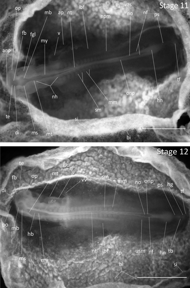
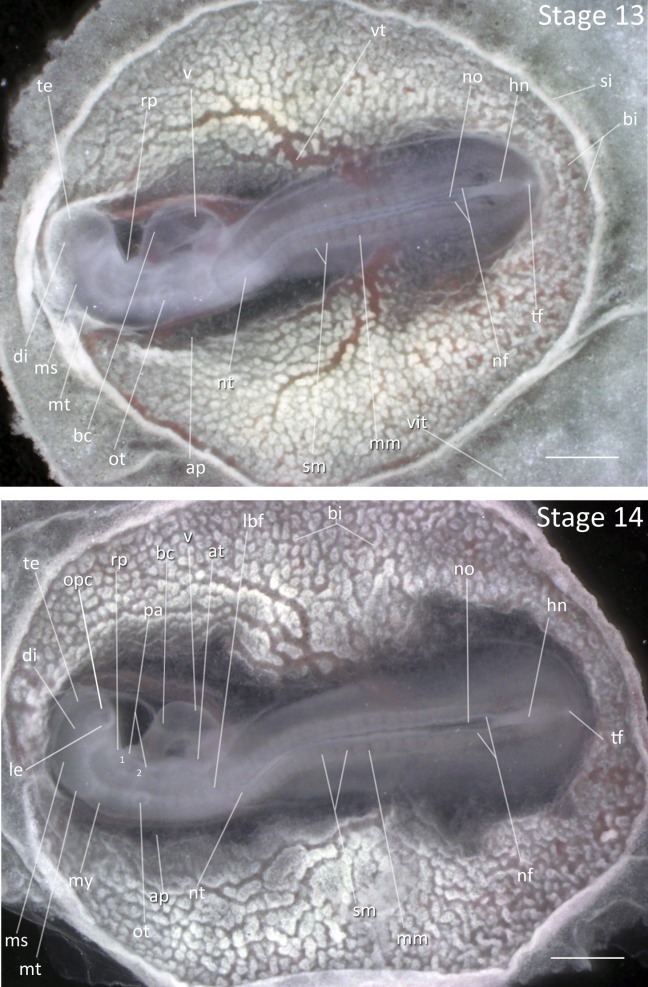
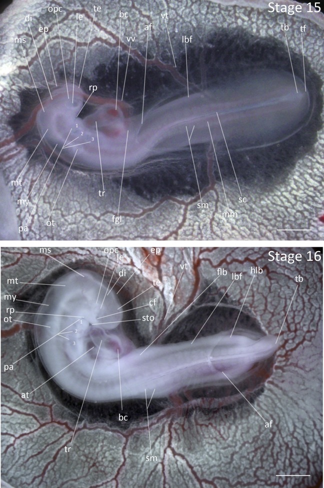
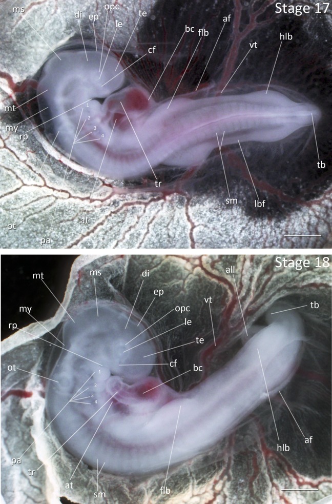
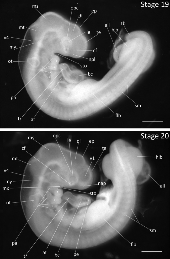
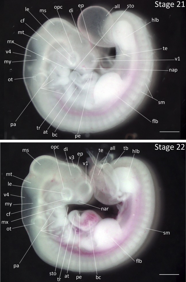
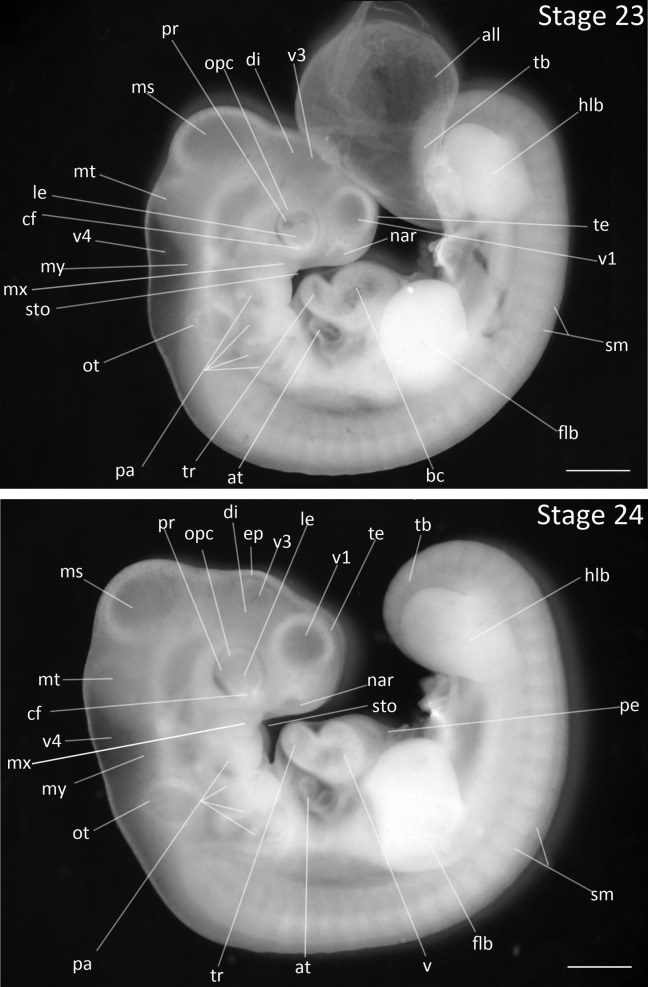
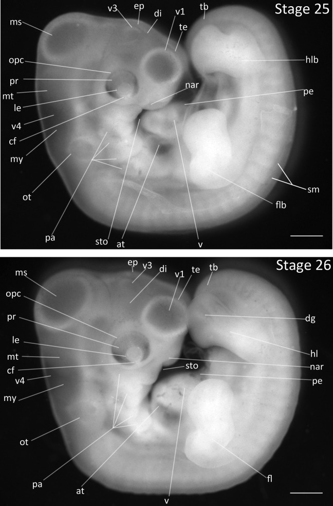
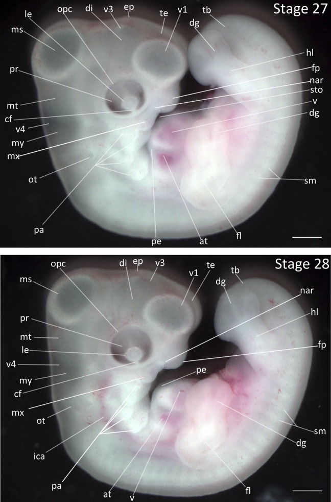
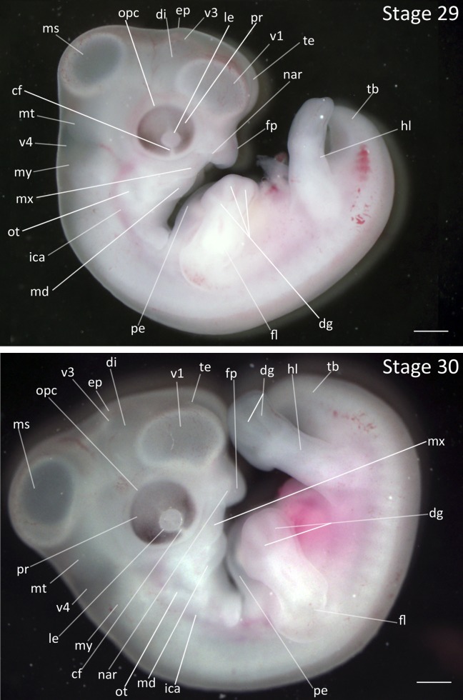
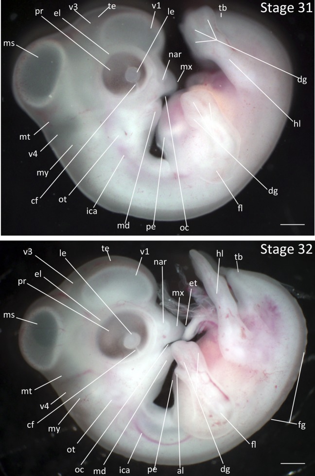
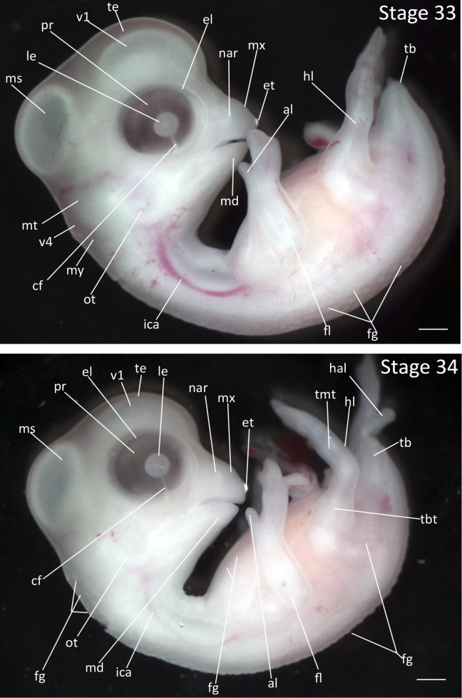
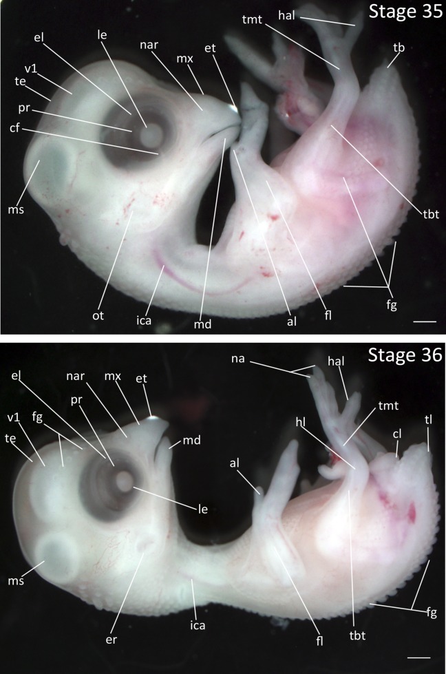
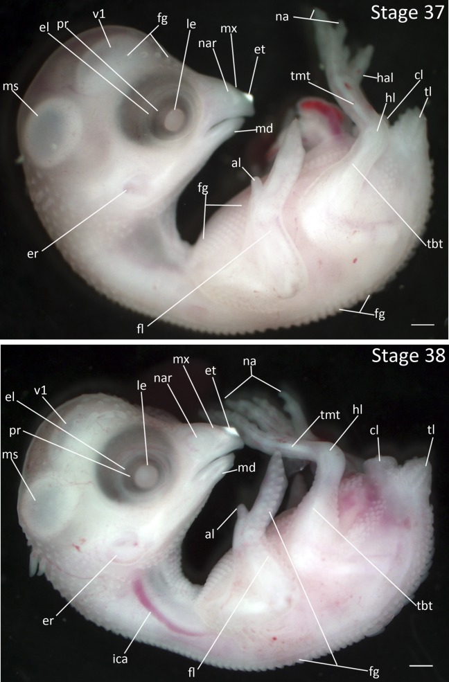
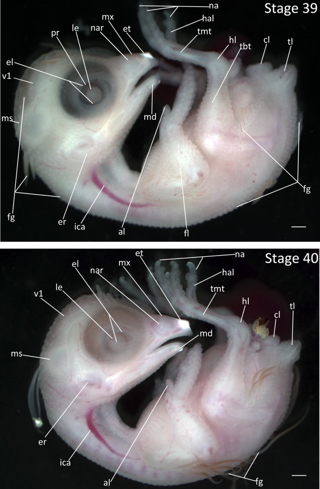
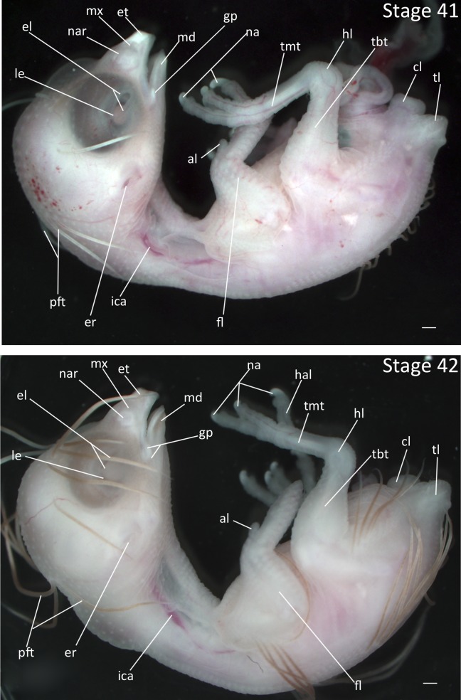
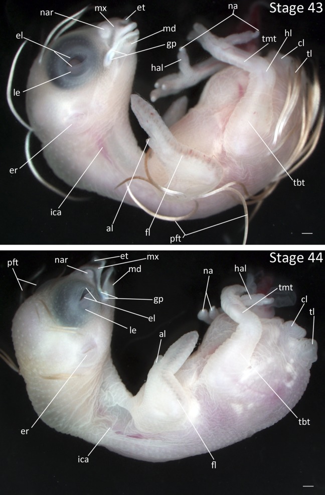
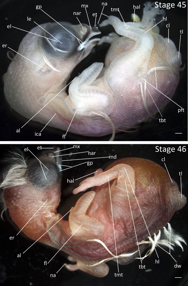
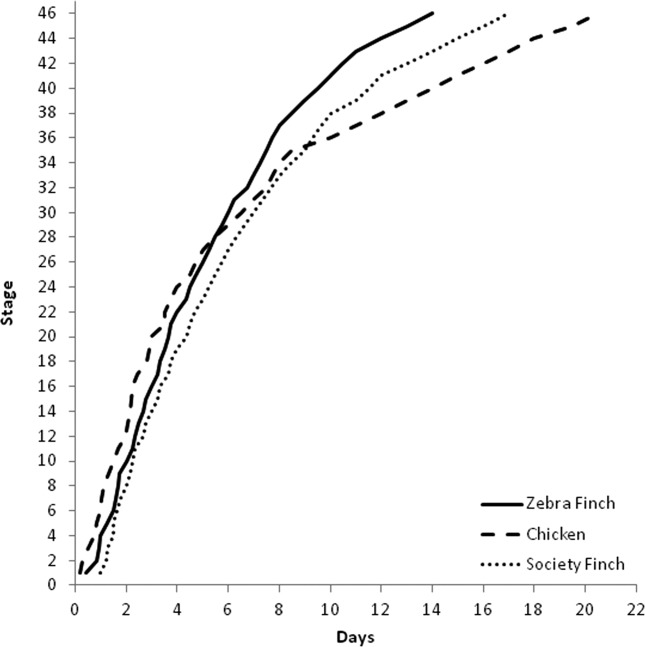
Zebra Finches (Taeniopygia guttata) are the most commonly used laboratory songbird species, yet their embryological development has been poorly characterized. Most studies to date apply Hamburger and Hamilton stages derived from chicken development; however, significant differences in development between precocial and altricial species suggest that they may not be directly comparable. We provide the first detailed description of embryological development in the Zebra Finch under standard artificial incubation. These descriptions confirm that some of the features used to classify chicken embryos into stages are not applicable in an altricial bird such as the Zebra Finch. This staging protocol will help to standardize future studies of embryological development in the Zebra Finch.

Keywords: Zebra Finch; artificial incubation; embryological development; passerine bird; staging.


Figures

Figure 1
Figure 1
Stages 1–4: All embryos oriented with anterior region at top of image. Scale bar = 1 mm. Stage 1 (0–20 h). Stage 2 (20–22 h). Stage 3 (22 h). Stage 4 (24 h). Abbreviations: af, amniotic fold; am, anterior margin of mesoderm; ao, area opaca; ap, area pellucida; es, embryonic shield; pf, primitive fold, lateral lip; pg, primitive groove, blastopore; ps, primitive streak; ta, thickened area; zj, zone of junction.
Figure 2
Figure 2
Stages 5 and 6: Anterior–posterior axis reads left to right in both images. Scale bar = 1 mm. Stage 5 (30 h). Stage 6 (36 h). Abbreviations: am, anterior margin of mesoderm; anp, anterior neural pore; ap, area pellucida; hn, Hensen’s node; hp, head process; mfh, margin of free head; nf, neural fold; no, notochord, notochord mesoderm; pf, primitive fold, lateral lip; pg, primitive groove, blastopore; po, proamnion, anterior blastopore; ps, primitive streak; zj, zone of junction. [Color figure can be viewed in the online issue, which is available at wileyonlinelibrary.com.]
Figure 3
Figure 3
Stages 7 and 8: Anterior–posterior axis reads left to right in both images. Scale bar = 1 mm. Stage 7 (38 h). Two somites. Embryos may have one to three somites visible. Stage 8 (40 h). Four to six somites. Abbreviations: aip, anterior intestinal portal; am, anterior margin of mesoderm; anp, anterior neural pore; ap, area pellucida; bi, blood islands; hf, head fold; hm, head mesoderm; hn, Hensen’s node; lbf, lateral body fold; mfh, margin of free head; mm, mesomere, nephrotome, intermediate mesoderm; nf, neural fold; no, notochord, notochord mesoderm; nt, neural tube; onp, open neural plate; po, proamnion; ps, primitive streak; si, sinus terminalis (circular vein of yolk sac); sm, somite (epimere) mesoderm; usm, unsegmented somite mesoderm; vas, area vasculosa; vit, area vitellina.
Figure 4
Figure 4
Stages 9 and 10: Anterior–posterior axis reads left to right in both images. Scale bar = 1 mm. Stage 9 (42 h). Seven somites. Stage 10 (2 days). 10 somites. Abbreviations: aip, anterior intestinal portal; amm, anterior margin of invaginated lateral mesoderm; anp, anterior neural pore; ao, area opaca; ap, area pellucida; bi, blood islands; fb, forebrain; fgl, foregut lumen; hb, hindbrain; hm, head mesoderm; hn, Hensen’s node; lbf, lateral body fold mesoderm; mb, midbrain; mfh, margin of free head; mm, mesomere; nf, neural fold; nh, rhombomeres of hindbrain; no, notochord; nt, neural tube; onp, open neural plate; op, optic vesicle; ps, primitive streak; si, sinus terminalis; sm, somite mesoderm; usm, unsegmented somite mesoderm; v, ventricle of heart; vas, area vasculosa; vit, area vitellina; vv, vitelline vein; zj, zone of junction.
Figure 5
Figure 5
Stages 11 and 12: Anterior–posterior axis reads left to right in both images. Scale bar = 1 mm. Stage 11 (2 ¼ days). Stage 12 (2 ⅓ days). Abbreviations: anp, anterior neural pore; ap, area pellucida; bc, bulbus cordis; bi, blood islands; di, diencephalon; fb, forebrain; fgl, foregut lumen; hb, hindbrain; hg, hindgut lumen; hn, Hensen’s node; lbf, lateral body fold mesoderm; mb, midbrain; mm, mesomere; ms, mesencephalon; mt, metencephalon; my, myelencephalon; nf, neural fold; nh, rhombomeres of hindbrain; no, notochord; npm, nephrogenic mesoderm; nt, neural tube; onp, open neural plate; op, optic vesicle; po, proamnion; ps, primitive streak; si, sinus terminalis; sm, somite mesoderm; tb, tailbud; te, telencephalon; tf, tail fold; usm, unsegmented somite mesoderm; v, ventricle of heart; vas, area vasculosa; vv, vitelline vein.
Figure 6
Figure 6
Stages 13 and 14: Anterior–posterior axis reads left to right in both images. Scale bar = 1 mm. Stage 13 (2 ½ days). Stage 14 (2 ⅔ days). Abbreviations: ap, area pellucida; at, atrium of heart; bc, bulbus cordis; bi, blood islands; di, diencephalon; hn, Hensen’s node; lbf, lateral body of foregut; le, lens vesicle; mm, mesomere; ms, mesencephalon; mt, metencephalon; my, myelencephalon; nf, neural fold; no, notochord; nt, neural tube; opc, optic cup; ot, otic vesicle; pa, pharyngeal arch; rp, Rathke’s pouch; si, sinus terminalis; sm, somite mesoderm; te, telencephalon; tf, tail fold; v, ventricle of heart; vit, area vitellina; vt, vitelline artery. [Color figure can be viewed in the online issue, which is available at wileyonlinelibrary.com.]
Figure 7
Figure 7
Stages 15 and 16: Anterior–posterior axis reads left to right in both images. Scale bar = 1 mm. Stage 15 (2 ¾ days). Stage 16 (3 days). Abbreviations: af, amniotic fold; at, atrium of heart; bc, bulbus cordis; cf, choroid fissure; di, diencephalon; ep, epiphysis; fgl, foregut lumen; flb, forelimb bud; hlb, hindlimb bud; lbf, lateral body of foregut; le, lens vesicle; mm, mesomere; ms, mesencephalon; mt, metencephalon; my, myelencephalon; opc, optic cup; ot, otic vesicle; pa, pharyngeal arch; rp, Rathke’s pouch; sc, spinal cord; sm, somite mesoderm; sto, stomodeum; tb, tailbud; te, telencephalon; tf, tail fold; tr, truncus arteriosus; vt, vitelline artery; vv, vitelline vein. [Color figure can be viewed in the online issue, which is available at wileyonlinelibrary.com.]
Figure 8
Figure 8
Stages 17 and 18: Anterior–posterior axis reads left to right in both images. Scale bar = 1 mm. Stage 17 (3 ¼ days). Stage 18 (3 ⅓ days). Abbreviations: af, amniotic fold; all, allantois; at, atrium of heart; bc, bulbus cordis; cf, choroid fissure; di, dienchepalon; ep, epiphysis; flb, forelimb bud; hlb, hindlimb bud; lbf, lateral body of foregut; le, lens vesicle; ms, mesencephalon; mt, metencephalon; my, myelencephalon; opc, optic cup; ot, otic vesicle; pa, pharyngeal arch; rp, Rathke’s pouch; sm, somite mesoderm; tb, tailbud; te, telencephalon; tr, truncus arteriosus; vt, vitelline artery. [Color figure can be viewed in the online issue, which is available at wileyonlinelibrary.com.]
Figure 9
Figure 9
Stages 19 and 20: Anterior–posterior axis reads left to right in both images. Scale bar = 1 mm. Stage 19 (3 ½ days). Stage 20 (3 ⅔ days). Abbreviations: all, allantois; at, atrium; bc, bulbus cordis; cf, choroid fissure; di, diencephalon; ep, epiphysis; flb, forelimb bud; hlb, hindlimb bud; le, lens vesicle; ms, mesencephalon; mt, metencephalon; my, myelencephalon; nap, nasal pit; npl, nasal placode; opc, optic cup; ot, otic vesicle; pa, pharyngeal arch; pe, pericardium; sm, somite mesoderm; sto, stomodeum; tb, tailbud; te, telencephalon; tr, truncus arteriosus; v1, lateral ventricle of telencephalon; v4, fourth vesicle of myelencephalon.
Figure 10
Figure 10
Stages 21 and 22: Anterior–posterior axis reads left to right in both images. Scale bar = 1 mm. Stage 21 (3 ¾ days). Stage 22 (4 days). Abbreviations: all, allantois; at, atrium; bc, bulbus cordis; cf, choroid fissure; di, diencephalon; ep, epiphysis; flb, forelimb bud; hlb, hindlimb bud; le, lens vesicle; ms, mesencephalon; mt, metencephalon; my, myelencephalon; nap, nasal pit; nar, nares; opc, optic cup; ot, otic vesicle; pa, pharyngeal arch; pe, pericardium; sm, somite mesoderm; sto, stomodeum; tb, tailbud; te, telencephalon; tr, truncus arteriosus; v1, lateral ventricle of telencephalon; v3, third ventricle of diencephalon; v4, fourth vesicle of myelencephalon. [Color figure can be viewed in the online issue, which is available at wileyonlinelibrary.com.]
Figure 11
Figure 11
Stages 23 and 24: Anterior–posterior axis reads left to right in both images. Scale bar = 1 mm. Stage 23 (4 ⅓ days). Stage 24 (4 ½ days). Abbreviations: all, allantois; at, atrium; bc, bulbus cordis; cf, choroid fissure; di, diencephalon; ep, epiphysis; flb, forelimb bud; hlb, hindlimb bud; le, lens vesicle; ms, mesencephalon; mt, metencephalon; my, myelencephalon; nar, nares; opc, optic cup; ot, otic vesicle; pa, pharyngeal arch; pe, pericardium; sm, somite mesoderm; sto, stomodeum; tb, tailbud; te, telencephalon; tr, truncus arteriosus; v, ventricle of heart; v1, lateral ventricle of telencephalon; v3, third ventricle of diencephalon; v4, fourth vesicle of myelencephalon.
Figure 12
Figure 12
Stages 25 and 26: Anterior–posterior axis reads left to right in both images. Scale bar = 1 mm. Stage 25 (4 ¾ days). Stage 26 (5 days). Abbreviations: all, allantois; at, atrium; cf, choroid fissure; dg, digit of limb; di, diencephalon; ep, epiphysis; fl, forelimb; flb, forelimb bud; hl, hindlimb; hlb, hindlimb bud; le, lens vesicle; ms, mesencephalon; mt, metencephalon; my, myelencephalon; nar, nares; opc, optic cup; ot, otic vesicle; pa, pharyngeal arch; pe, pericardium; sm, somite mesoderm; sto, stomodeum; tb, tailbud; te, telencephalon; v, ventricle of heart; v1, lateral ventricle of telencephalon; v3, third ventricle of diencephalon; v4, fourth vesicle of myelencephalon.
Figure 13
Figure 13
Stages 27 and 28: Anterior–posterior axis reads left to right in both images. Scale bar = 1 mm. Stage 27 (5 ¼ days). Stage 28 (5 ½ days). Abbreviations: all, allantois; at, atrium; cf, choroid fissure; dg, digit of limb; di, diencephalon; ep, epiphysis; fl, forelimb; fp, frontal process; hl, hindlimb; ica, internal carotid artery; le, lens vesicle; ms, mesencephalon; mt, metencephalon; mx, maxillary process; my, myelencephalon; nar, nares; opc, optic cup; ot, otic vesicle; pa, pharyngeal arch; pe, pericardium; sm, somite mesoderm; sto, stomodeum; tb, tailbud; te, telencephalon; v, ventricle of heart; v1, lateral ventricle of telencephalon; v3, third ventricle of diencephalon; v4, fourth vesicle of myelencephalon. [Color figure can be viewed in the online issue, which is available at wileyonlinelibrary.com.]
Figure 14
Figure 14
Stages 29 and 30: Anterior–posterior axis reads left to right in both images. Scale bar = 1 mm. Stage 29 (5 ¾ days). Stage 30. (6 days). Abbreviations: all, allantois; cf, choroid fissure; dg, digit of limb; di, diencephalon; ep, epiphysis; fl, forelimb; fp, frontal process; hl, hindlimb; ica, internal carotid artery; le, lens vesicle; md, mandibular process; ms, mesencephalon; mt, metencephalon; mx, maxillary process; my, myelencephalon; nar, nares; opc, optic cup; ot, otic vesicle; pe, pericardium; sm, somite mesoderm; sto, stomodeum; tb, tailbud; te, telencephalon; v1, lateral ventricle of telencephalon; v3, third ventricle of diencephalon; v4, fourth vesicle of myelencephalon. [Color figure can be viewed in the online issue, which is available at wileyonlinelibrary.com.]
Figure 15
Figure 15
Stages 31 and 32: Anterior–posterior axis reads left to right in both images. Scale bar = 1 mm. Stage 31 (6 ¼ days). Stage 32 (6 ¾ days). Abbreviations: all, allantois; cf, choroid fissure; dg, digit of limb; di, diencephalon; el, eyelid; ep, epiphysis; fg, feather germ; fl, forelimb; hl, hindlimb; ica, internal carotid artery; le, lens vesicle; md, mandibular process; ms, mesencephalon; mt, metencephalon; mx, maxillary process; my, myelencephalon; nar, nares; opc, optic cup; ot, otic vesicle; pe, pericardium; sm, somite mesoderm; sto, stomodeum; tb, tailbud; te, telencephalon; v1, lateral ventricle of telencephalon; v3, third ventricle of diencephalon; v4, fourth vesicle of myelencephalon. [Color figure can be viewed in the online issue, which is available at wileyonlinelibrary.com.]
Figure 16
Figure 16
Stages 33 and 34: Anterior–posterior axis reads left to right in both images. Scale bar = 1 mm. Stage 33 (7 days). Stage 34 (7 ¼ days). Abbreviations: al, alula; cf, choroid fissure; el, eyelid; et, egg tooth; fg, feather germ; fl, forelimb; hal, hallux; hl, hindlimb; ica, internal carotid artery; le, lens vesicle; md, mandibular process; ms, mesencephalon; mt, metencephalon; mx, maxillary process; my, myelencephalon; nar, nares; ot, otic vesicle; pr, pigmented retina of eye; tb, tailbud; te, telencephalon; tmt, tarsometatarsus; v1, lateral ventricle of telencephalon; v4, fourth vesicle of myelencephalon. [Color figure can be viewed in the online issue, which is available at wileyonlinelibrary.com.]
Figure 17
Figure 17
Stages 35 and 36: Anterior–posterior axis reads left to right in both images. Scale bar = 1 mm. Stage 35 (7 ½ days). Stage 36 (7 ¾ days). Abbreviations: al, alula; cf, choroid fissure; el, eyelid; er, opening to ear; et, egg tooth; fg, feather germ; fl, forelimb; hal, hallux; hl, hindlimb; ica, internal carotid artery; le, lens vesicle; md, mandibular process; ms, mesencephalon; mx, maxillary process; na, nail; nar, nares; ot, otic vesicle; pr, pigmented retina of eye; tb, tailbud; tbt, tibiotarsus; te, telencephalon; tl, tail; tmt, tarsometatarsus; v1, lateral ventricle of telencephalon. [Color figure can be viewed in the online issue, which is available at wileyonlinelibrary.com.]
Figure 18
Figure 18
Stages 37 and 38: Anterior–posterior axis reads left to right in both images. Scale bar = 1 mm. Stage 37 (8 days). Stage 38 (8 ½ days). Abbreviations: al, alula; cl, cloaca; el, eyelid; er, opening to ear; et, egg tooth; fg, feather germ; fl, forelimb; hal, hallux; hl, hindlimb; ica, internal carotid artery; le, lens vesicle; md, mandibular process; ms, mesencephalon; mx, maxillary process; na, nail; nar, nares; pr, pigmented retina of eye; tbt, tibiotarsus; tl, tail; tmt, tarsometatarsus; v1, lateral ventricle of telencephalon. [Color figure can be viewed in the online issue, which is available at wileyonlinelibrary.com.]
Figure 19
Figure 19
Stages 39 and 40: Anterior–posterior axis reads left to right in both images. Scale bar = 1 mm. Stage 39 (9 days). Stage 40 (9 ½ days). Anterior–posterior axis reads left to right. Abbreviations: al, alula; cl, cloaca; el, eyelid; er, opening to ear; et, egg tooth; fg, feather germ; fl, forelimb; hal, hallux; hl, hindlimb; ica, internal carotid artery; le, lens vesicle; md, mandibular process; ms, mesencephalon; mx, maxillary process; na, nail; nar, nares; pr, pigmented retina of eye; tbt, tibiotarsus; tl, tail; tmt, tarsometatarsus; v1, lateral ventricle of telencephalon. [Color figure can be viewed in the online issue, which is available at wileyonlinelibrary.com.]
Figure 20
Figure 20
Stages 41 and 42: Anterior–posterior axis reads left to right in both images. Scale bar = 1 mm. Stage 41 (10 days). Stage 42 (10 ½ days). Abbreviations: al, alula; cl, cloaca; el, eyelid; er, opening to ear; et, egg tooth; fl, forelimb; gp, gape flange; hal, hallux; hl, hindlimb; ica, internal carotid artery; le, lens vesicle; md, mandibular process; mx, maxillary process; na, nail; nar, nares; pft, pin feathers; tbt, tibiotarsus; tl, tail; tmt, tarsometatarsus. [Color figure can be viewed in the online issue, which is available at wileyonlinelibrary.com.]
Figure 21
Figure 21
Stages 43 and 44: Anterior–posterior axis reads left to right in both images. Scale bar = 1 mm. Stage 43 (11 days). Stage 44 (12 days). Abbreviations: al, alula; cl, cloaca; el, eyelid; er, opening to ear; et, egg tooth; fl, forelimb; gp, gape flange; hal, hallux; hl, hindlimb; ica, internal carotid artery; le, lens vesicle; md, mandibular process; mx, maxillary process; na, nail; nar, nares; pft, pin feathers; tbt, tibiotarsus; tl, tail; tmt, tarsometatarsus. [Color figure can be viewed in the online issue, which is available at wileyonlinelibrary.com.]
Figure 22
Figure 22
Stages 45 and 46: Anterior–posterior axis reads left to right in both images. Scale bar = 1 mm. Stage 45 (13 days). Stage 46 (14 days). Abbreviations: al, alula; cl, cloaca; dw, down feathers; el, eyelid; er, opening to ear; et, egg tooth; fl, forelimb; gp, gape flange; hal, hallux; hl, hindlimb; ica, internal carotid artery; le, lens vesicle; md, mandibular process; mx, maxillary process; na, nail; nar, nares; pft, pin feathers; tbt, tibiotarsus; tl, tail; tmt, tarsometatarsus. [Color figure can be viewed in the online issue, which is available at wileyonlinelibrary.com.]
Figure 23
Figure 23
Timing of the stages in the Zebra Finch, Society Finch, and chicken. Solid line is the development of the Zebra Finch, dashed line is the development of the chicken, and dotted line is the development of Society Finch. The timing of the stages is determined from the average time reported in staging papers (Hamburger and Hamilton, ; Yamasaki and Tonosaki, 1988).

References

    1. Austad SN. Candidate bird species for use in aging research. ILAR J. 2011;52:89–96. - PubMed
    1. Bird DM, Gautier J, Montpetit V. Embryonic growth of American Kestrels. Auk. 1984;101:392–396.
    1. Blom J, Lilja C. A comparative study of embryonic development of some bird species with different patterns of postnatal growth. Zoology. 2005;108:81–95. - PubMed
    1. Bottjer SW, To M. Afferents from vocal motor and respiratory effectors are recruited during vocal production in juvenile songbirds. J Neurosci. 2012;32:10895–10906. - PMC - PubMed
    1. Chen C-C, Balaban E, Jarvis ED. 2012. Interspecies avian brain chimeras reveal that large brain size differences are influenced by cell-interdependent processes. PLoS One 7:e42477. - PMC - PubMed

Publication types

LinkOut - more resources