. 2013 Sep 1;126(Pt 17):3904-15.
doi: 10.1242/jcs.126565. Epub 2013 Jun 26.

ZO-1 recruitment to α-catenin--a novel mechanism for coupling the assembly of tight junctions to adherens junctions

Affiliations

ZO-1 recruitment to α-catenin--a novel mechanism for coupling the assembly of tight junctions to adherens junctions

Jessica L Maiers et al. J Cell Sci. .

Abstract

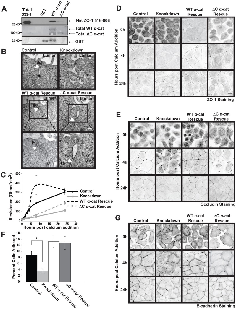
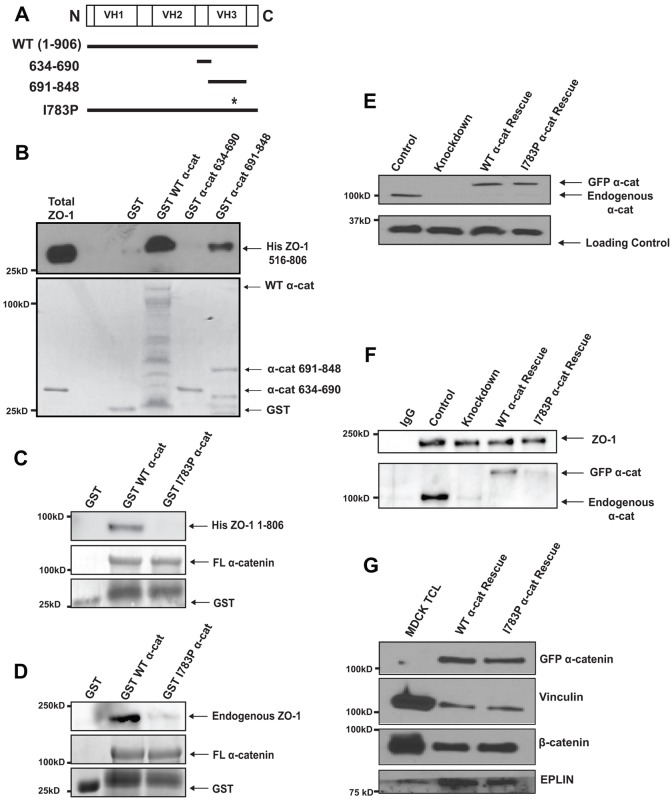
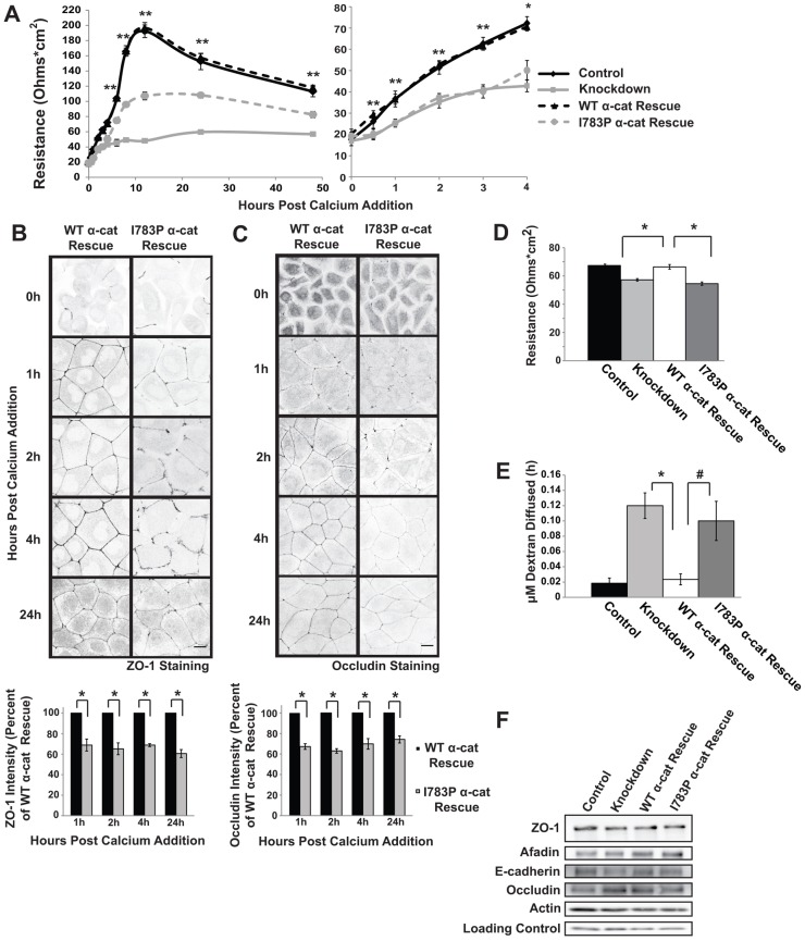
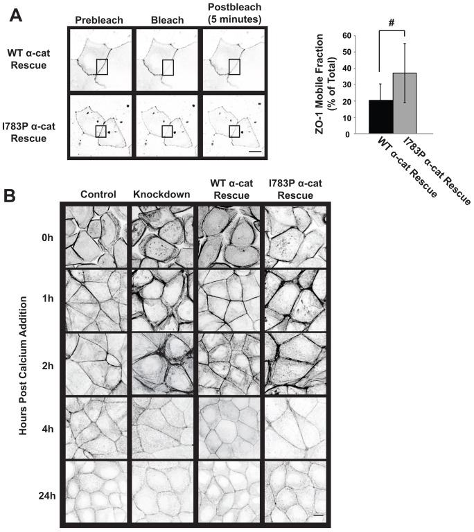
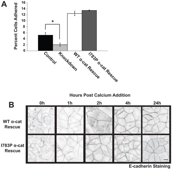
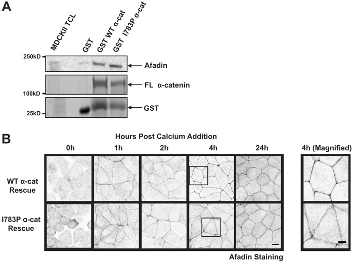
The formation of a barrier between epithelial cells is a fundamental determinant of cellular homeostasis, protecting underlying cells against pathogens, dehydration and damage. Assembly of the tight junction barrier is dependent upon neighboring epithelial cells binding to one another and forming adherens junctions, but the mechanism for how these processes are linked is poorly understood. Using a knockdown and substitution system, we studied whether ZO-1 binding to α-catenin is required for coupling tight junction assembly to the formation of adherens junctions. We found that preventing ZO-1 binding to α-catenin did not appear to affect adherens junctions. Rather the assembly and maintenance of the epithelial barrier were disrupted. This disruption was accompanied by alterations in the mobility of ZO-1 and the organization of the actin cytoskeleton. Thus, our study identifies α-catenin binding to ZO-1 as a new mechanism for coupling the assembly of the epithelial barrier to cell-to-cell adhesion.

Keywords: Adherens junction; Tight junction; ZO-1; α-Catenin.


Figures

Fig. 1.
Fig. 1.
The α-catenin C-terminus is required for the assembly of tight junctions, but not adherens junctions. (A) The α-catenin C-terminus is necessary for ZO-1 binding. GST or GST fused to either a full-length α-catenin or a C-terminal-truncated form of α-catenin were purified, bound to glutathione–Sepharose beads and incubated with a purified fragment of ZO-1 containing the predicted α-catenin-binding site (residues 516–806). The proteins were recovered, resolved using SDS-PAGE and immunoblotted with an antibody that recognizes the His tag on ZO-1 (upper panel). The immunoblot was stained with Coomassie Blue to show the levels of the GST proteins employed in the assay (lower panels). (B) Tight junctions and adherens junctions display no gross structural alterations in ΔC α-cat Rescue cells. MDCK II cells were infected with GFP-tagged α-catenin (WT α-cat Rescue) or the C-terminal truncation mutant fused to GFP (ΔC α-cat Rescue) and infected a second time with viruses encoding shRNAs targeting canine α-catenin (Knockdown). Cells expressing GFP and an empty shRNA targeting vector were used as a control (Control). The cell–cell junctions were visualized using TEM 4 hours after the assembly of cell–cell junctions was initiated by restoring Ca2+ to Ca2+-starved cells. The black arrows denote tight junctions and white arrows denote adherens junctions. Insets of the boxed areas within WT α-cat Rescue and ΔC α-cat Rescue micrographs are located below the original images. Scale bars: 0.2 µm for top 4 images (shown in the middle row), 0.1 µm for insets. (C) The permeability of the epithelial monolayer is disrupted during junction assembly in ΔC α-cat Rescue cells. The transelectrical epithelial resistance (TER) in confluent cultures of cells was measured using a voltometer at 0, 1, 2, 4, 6, 8 and 24 hours after junctional assembly was initiated by restoring Ca2+ to serum-starved cells. The data in the graph represent the mean±s.e.m. for four independent experiments. (D,E) The integrity of the tight junctions is altered in ΔC α-cat Rescue cells. ZO-1 (D) and occludin (E) localization was examined by immunofluorescence in Control, Knockdown, WT α-cat Rescue and ΔC α-cat Rescue cells that had been incubated in Ca2+-depleted medium (0 h) 4 or 24 hours after Ca2+ restoration. Representative confocal images are shown in inverted grayscale so that the differences between WT α-cat Rescue and ΔC α-cat Rescue cells can be readily visualized. Scale bar: 10 µm. (F) Cadherin-mediated adhesion is maintained in ΔC α-cat Rescue cells. Control, Knockdown, WT α-cat Rescue and ΔC α-cat Rescue cell lines were plated on surfaces coated with cadherin extracellular domains, washed, and the adherent cells were counted. The data presented in the graph are the mean±s.e.m. from four independent experiments. *P≤0.01 (G) ΔC α-cat Rescue cells display proper localization of E-cadherin. E-cadherin localization was examined by immunofluorescence in Control, Knockdown, WT α-cat Rescue or ΔC α-cat Rescue cells incubated overnight in Ca2+-depleted medium (0 h), 4 or 24 hours after Ca2+ was restored to the cell cultures. Representative pictures are shown in inverted grayscale. Scale bar: 10 µm.
Fig. 2.
Fig. 2.
α-Catenin harboring an I783P substitution does not bind ZO-1 in vitro or in MDCKII cells. (A) A linear schematic of α-catenin and the fragments of α-catenin used in this study (GST–α-catenin). (B,C) In vitro binding of ZO-1 to various GST–α-catenin proteins. The indicated α-catenin proteins were purified, attached to beads and incubated with purified His-tagged ZO-1 (516–806 for B; 1–806 for C). The levels of GST-tagged α-catenin proteins employed were similar, as represented by the Coomassie-stained blot in the lower panels. Note that in C, ZO-1 fails to bind α-catenin I783P. We consistently obtained some GST in the preparations of the full-length (FL) α-catenin proteins. (D) Endogenous ZO-1 binds WT α-catenin but not I783P α-catenin. GST, GST-tagged WT α-catenin or GST-tagged I783P α-catenin were prebound to glutathione beads and incubated with MDCKII cell lysates. The GST-tagged proteins were recovered and separated using SDS-PAGE. The co-precipitating levels of ZO-1 were examined by immunoblotting. This figure shows that endogenous, full-length ZO-1 binds to WT α-catenin, but fails to bind I783P α-catenin. (E) Expression levels of I783P α-catenin in MDCKII cells. MDCKII cells expressing an shRNA against canine α-catenin were infected a second time with retroviruses encoding GFP–α-catenin with the I783P substitution (I783P α-cat Rescue). Lysates were harvested from cell lines stably expressing these proteins or Control, Knockdown or WT α-cat Rescue cells. Immunoblot analysis was performed using antibodies against α-catenin or the p34-Arc subunit of the Arp2/3 complex as a loading control. (F) ZO-1 fails to co-immunoprecipitate with α-catenin I783P. Confluent monolayers of the indicated cell lines were Ca2+-starved overnight and lysed 1 hour post Ca2+ addition. ZO-1 was immunoprecipitated, and the bound proteins were washed, resolved using SDS-PAGE, and immunoblotted with an antibody against ZO-1 or α-catenin. (G) Substitution of I783P does not impair α-catenin binding to other ligands. WT α-cat Rescue or I783P α-cat Rescue cells were grown to confluence, lysed and full-length α-catenin and α-catenin I783P were immunoprecipitated using an antibody against GFP. Proteins were recovered, resolved using SDS-PAGE, and immunoblotted with antibodies against vinculin, β-catenin or EPLIN to show the levels of each protein recovered, or GFP to show the levels of each α-catenin protein recovered.
Fig. 3.
Fig. 3.
Tight junctions are altered in cells expressing α-catenin with the I783P substitution. (A) The establishment of an epithelial barrier is disrupted in cells expressing I783P α-catenin. Confluent cultures of the indicated cells lines were incubated overnight in Ca2+-free medium (0 h). Ca2+ was restored to the cultures for the indicated times and resistance was measured using a voltometer on quadruplicate filters. The graph displays the mean±s.d. expressed in Ohms*cm2. The right panel shows the early time points (0–4 h) so that the initial delay in barrier establishment can be visualized. *P≤0.01 and **P≤0.001 compared with control. (B,C) The integrity of the tight junction is altered in cells expressing the α-catenin I783P point mutant that doesn't bind ZO-1. WT α-cat Rescue and I783P α-cat Rescue cells were examined using immunofluorescence and antibodies against ZO-1 (B) or occludin (C). Representative confocal images are shown in inverted grayscale. Scale bars: 10 µm. The intensity of ZO-1 or occludin at cell–cell contacts was determined using ImageJ and is shown below the images as the percentage intensity compared with WT α-cat Rescue. *P≤0.01. (D,E) The maintenance of the epithelial barrier is disrupted in I783P-expressing cells. (D) The indicated cell lines were grown to confluence, and resistance across the cell monolayer was measured daily until a consistent reading was made for three consecutive days. The graphed data represents the mean±s.e.m. for four independent experiments. The paracellular flux of 3 kDa FITC-conjugated dextran in the same cultures was measured (E) and the graph represents the mean±s.e.m. for four independent experiments. #P≤0.05; *P≤0.01. (F) Expression of tight and adherens junction proteins are unaltered in I783P α-cat Rescue cells. Lysates from the indicated cell lines were harvested and immunoblotting was performed to analyze the expression levels of ZO-1, occludin, actin, afadin and E-cadherin. p34-Arc serves as a loading control.
Fig. 4.
Fig. 4.
ZO-1 mobility and actin organization is altered by proline substitution at I783. (A) ZO-1 mobility is increased in I783P α-cat Rescue cells. ZO-1 mobility at cell–cell contacts was examined using FRAP. Cells were transfected with mCherry-tagged ZO-1 and FRAP was performed on adjacent transfected cells. The images presented are a representation of cells prior to bleaching (Prebleach), immediately after bleaching (Bleach) and 5 minutes after bleaching (Postbleach). The box indicates the bleached area. The amount of ZO-1 that recovered was quantified and the mobile fraction of ZO-1 was calculated and is shown in the right panel. (B) Actin organization is disrupted in Knockdown and I783P α-cat Rescue cells. Cells were grown to confluence and incubated overnight in Ca2+-free medium. Ca2+ was restored for the indicated times and actin organization was analyzed using immunofluorescence. Representative images are presented in inverted grayscale. Scale bars: 10 µm.
Fig. 5.
Fig. 5.
The proline substitution at I783 has no effect on adherens junction assembly or cadherin-mediated adhesion. (A) Cadherin-mediated adhesion is established in the presence of I783P α-catenin. The adhesion of the indicated cell lines to immobilized cadherin extracellular domains was examined as described in Fig. 1F. The data presented in the graph are the mean±s.e.m. cells adhered from four independent experiments. (B) E-cadherin localization is unaffected by I783P substitution. WT α-cat Rescue and I783P α-cat Rescue cells were grown to confluence, incubated in Ca2+-free medium overnight, and placed in Ca2+-containing medium for 0, 1, 2, 4 or 24 hours. The cells were then fixed, and examined by immunofluorescence using a Zeiss 510 confocal microscope. Images are presented in inverted grayscale. Scale bar: 10 µm.
Fig. 6.
Fig. 6.
Afadin localization and binding to α-catenin is unaffected by I783P substitution. (A) The I783P substitution has no effect on afadin binding to α-catenin. Purified GST-tagged FL α-catenin or I783P α-catenin were bound to glutathione beads and incubated with MDCKII cell lysates. The beads were washed and the bound proteins were separated using SDS-PAGE. Afadin levels were determined using by immunoblotting and the levels of GST-tagged WT α-catenin and I783P α-catenin loaded were determined by Coomassie staining. (B) Afadin localization to adherens junction during assembly is not affected by expression of I783P α-catenin. Afadin localization to cell–cell junctions was examined before (0 h) or 1, 2, 4 or 24 h after Ca2+ was restored to serum-starved cultures. Images are shown in inverted grayscale. The right panel displays magnified inserts of the boxed areas within the WT α-cat Rescue and I783P α-cat Rescue images at 4 h. Scale bars: 10 µm (B, left panels); 5 µm (B, right panels).

References

    1. Abe K., Takeichi M. (2008). EPLIN mediates linkage of the cadherin catenin complex to F-actin and stabilizes the circumferential actin belt. Proc. Natl. Acad. Sci. USA 105, 13–19 10.1073/pnas.0710504105 - DOI - PMC - PubMed
    1. Aberle H., Butz S., Stappert J., Weissig H., Kemler R., Hoschuetzky H. (1994). Assembly of the cadherin-catenin complex in vitro with recombinant proteins. J. Cell Sci. 107, 3655–3663 - PubMed
    1. Ando-Akatsuka Y., Yonemura S., Itoh M., Furuse M., Tsukita S. (1999). Differential behavior of E-cadherin and occludin in their colocalization with ZO-1 during the establishment of epithelial cell polarity. J. Cell. Physiol. 179, 115–125 10.1002/(SICI)1097-4652(199905)179:2<115::AID-JCP1>3.0.CO;2-T - DOI - PubMed
    1. Asakura T., Nakanishi H., Sakisaka T., Takahashi K., Mandai K., Nishimura M., Sasaki T., Takai Y. (1999). Similar and differential behaviour between the nectin-afadin-ponsin and cadherin-catenin systems during the formation and disruption of the polarized junctional alignment in epithelial cells. Genes Cells 4, 573–581 10.1046/j.1365-2443.1999.00283.x - DOI - PubMed
    1. Capaldo C. T., Macara I. G. (2007). Depletion of E-cadherin disrupts establishment but not maintenance of cell junctions in Madin-Darby canine kidney epithelial cells. Mol. Biol. Cell 18, 189–200 10.1091/mbc.E06-05-0471 - DOI - PMC - PubMed

Publication types