. 2013 Sep;33(9):2130-6.
doi: 10.1161/ATVBAHA.113.301826. Epub 2013 Jun 27.

Endothelial cell sensing of flow direction

Affiliations

Endothelial cell sensing of flow direction

Chong Wang et al. Arterioscler Thromb Vasc Biol. 2013 Sep.

Abstract

Objective: Atherosclerosis-prone regions of arteries are characterized by complex flow patterns where the magnitude of shear stress is low and direction rapidly changes, termed disturbed flow. How endothelial cells sense flow direction and how it impacts inflammatory effects of disturbed flow are unknown. We therefore aimed to understand how endothelial cells respond to changes in flow direction.

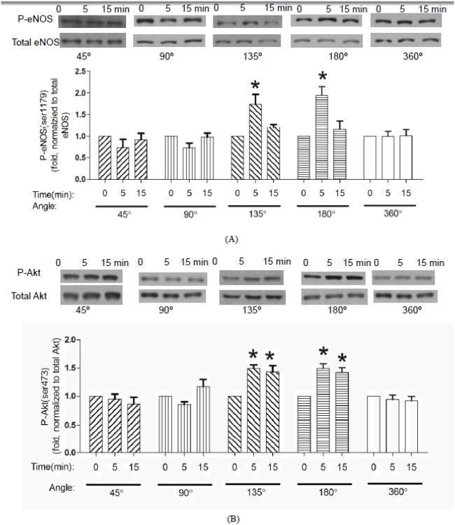
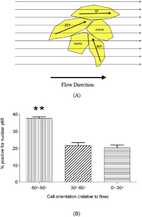
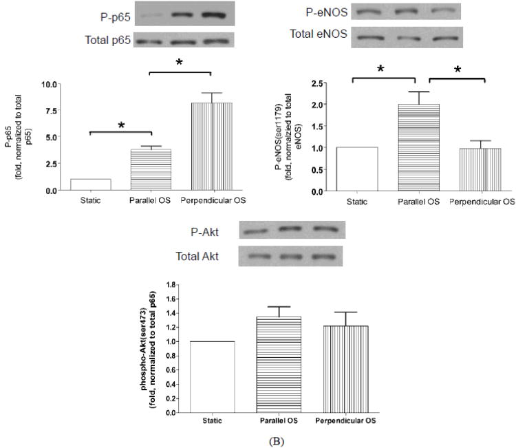
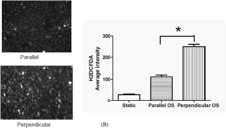
Approach and results: Using a recently developed flow system capable of changing flow direction to any angle, we show that responses of aligned endothelial cells are determined by flow direction relative to their morphological and cytoskeletal axis. Activation of the atheroprotective endothelial nitric oxide synthase pathway is maximal at 180° and undetectable at 90°, whereas activation of proinflammatory nuclear factor-κB is maximal at 90° and undetectable at 180°. Similar effects were observed in randomly oriented cells in naive monolayers subjected to onset of shear. Cells aligned on micropatterned substrates subjected to oscillatory flow were also examined. In this system, parallel flow preferentially activated endothelial nitric oxide synthase and production of nitric oxide, whereas perpendicular flow preferentially activated reactive oxygen production and nuclear factor-κB.

Conclusions: These data show that the angle between flow and the cell axis defined by their shape and cytoskeleton determines endothelial cell responses. The data also strongly suggest that the inability of cells to align in low and oscillatory flow is a key determinant of the resultant inflammatory activation.

Keywords: atherosclerosis; hemodynamics; mechanotransduction, cellular.


Figures

Figure 1
Figure 1
Activation of eNOS and Akt after changing flow direction. Cells aligned in flow for 24 h were subject to a change in direction by 45°, 90°, 135° and 180° and 360°. At the indicated times, cells were harvested and phospho-eNOS (ser1179) (A) and phospho-Akt (ser473) (B) were assayed by Western blotting. Values are means ± SEM, n=4; *P<0.05.
Figure 2
Figure 2
Activation of NF-kB after changing flow direction. Flow-aligned cells were subject to a change in flow direction as in Figure 1. Phospho-p65 was assayed by Western blotting. Values are means ± SEM, n=4; *P<0.05.
Figure 3
Figure 3
Responses of non-aligned cells to change of flow direction. Cells under flow for 2h were subject to a change in flow angle as in Figure 1. (A) Phospho-eNOS; (B) Phospho-p65; (C) Phospho-Akt. Values are means ± SEM, n=3.
Figure 4
Figure 4
Responses of naive cells to the onset of flow. (A): Cartoon of cells in a naive cell monolayer with different angles relative to the flow direction. (B): NF-κB nuclear translocation in elongated cells that were perpendicular (60°-90°), intermediate (30°-60°) or parallel (0°-30°) to the flow direction. Shear stress of 15 dynes/cm2 was applied for 30 min. Values are means ± SEM, n=3; **P<0.001.
Figure 5
Figure 5
Response of micropattern-aligned cells to oscillatory flow. Cells aligned on micropattterned fibronectin lines were analyzed by staining for p65 and visualizing nuclear translocation. (A): Images of cells without flow (left), and with 2h oscillatory flow parallel (center) or perpendicular (right) to the lines. (B): Cells were extracted and analyzed by Western blotting for phospho-p65 (left), phospho-eNOS (right) and phospho-Akt (bottom). Values are means ± SEM, n=3; *P<0.05.
Figure 5
Figure 5
Response of micropattern-aligned cells to oscillatory flow. Cells aligned on micropattterned fibronectin lines were analyzed by staining for p65 and visualizing nuclear translocation. (A): Images of cells without flow (left), and with 2h oscillatory flow parallel (center) or perpendicular (right) to the lines. (B): Cells were extracted and analyzed by Western blotting for phospho-p65 (left), phospho-eNOS (right) and phospho-Akt (bottom). Values are means ± SEM, n=3; *P<0.05.
Figure 6
Figure 6
Reactive oxygen and nitric oxide production. Cells on micropatterned surfaces were subject to oscillatory flow for 1h. (A): DAF-FM detection of nitric oxide. (B): H2DCFDA detection of ROS. Images on the left, quantified values on the right are means ± SEM, n=3; *P<0.05.
Figure 6
Figure 6
Reactive oxygen and nitric oxide production. Cells on micropatterned surfaces were subject to oscillatory flow for 1h. (A): DAF-FM detection of nitric oxide. (B): H2DCFDA detection of ROS. Images on the left, quantified values on the right are means ± SEM, n=3; *P<0.05.

References

    1. Berk B. Atheroprotective Signaling Mechanisms Activated by Steady Laminar Flow in Endothelial Cells. Circulation. 2008;117:1082–1089. - PubMed
    1. Chatzizisis Y, Coskun A, Jonas M, Edelman E, Feldman C, Stone P. Role of endothelial shear stress in the natural history of coronary atherosclerosis and vascular remodelingmolecular, cellular, and vascular behavior. Journal of the American College of Cardiology. 2007;49:2379–2393. - PubMed
    1. Chiu J, Chien S. Effects of disturbed flow on vascular endothelium: pathophysiological basis and clinical perspectives. Physiological reviews. 2011;91:327–387. - PMC - PubMed
    1. Davies P. Hemodynamic shear stress and the endothelium in cardiovascular pathophysiology. Nature Clinical Practice Cardiovascular Medicine. 2008;6:16–26. - PMC - PubMed
    1. Malek A, Alper S, Izumo S. Hemodynamic shear stress and its role in atherosclerosis. JAMA: The Journal of the American Medical Association. 1999;282:2035–2042. - PubMed

Publication types

MeSH terms