. 2013 Jul;5(4):216-23.
doi: 10.4168/aair.2013.5.4.216. Epub 2013 May 27.

Rhinovirus-Infected Epithelial Cells Produce More IL-8 and RANTES Compared With Other Respiratory Viruses

Affiliations

Rhinovirus-Infected Epithelial Cells Produce More IL-8 and RANTES Compared With Other Respiratory Viruses

Yoon Hong Chun et al. Allergy Asthma Immunol Res. 2013 Jul.

Abstract

Purpose: The environmental factors human rhinoviruses (HRVs) and house dust mites (HDMs) are the most common causes of acute exacerbations of asthma. The aim of this study was to compare the chemokine production induced by HRVs in airway epithelial cells with that induced by other respiratory viruses, and to investigate synergistic interactions between HRVs and HDMs on the induction of inflammatory chemokines in vitro.

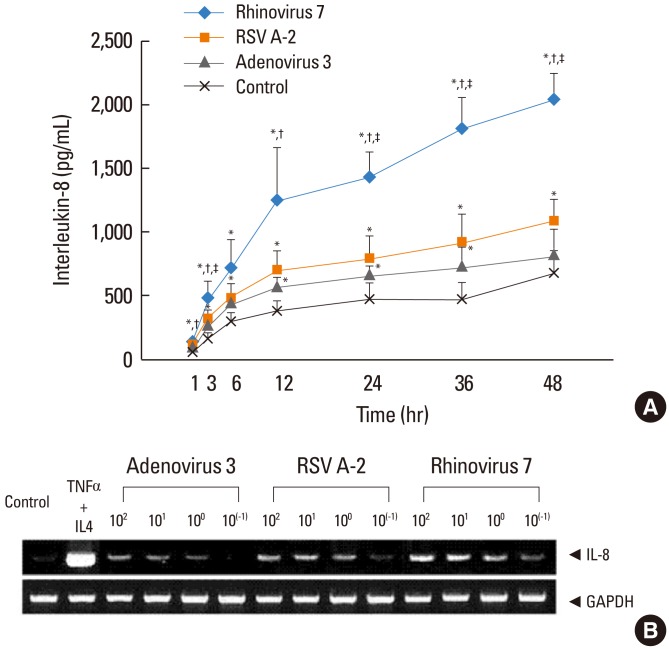
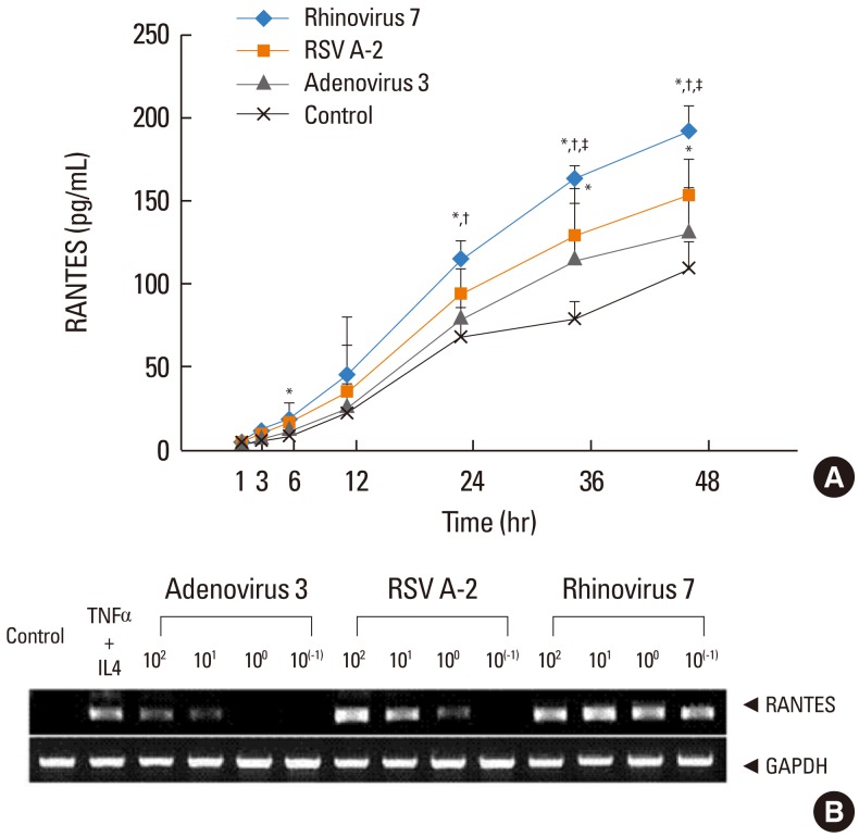
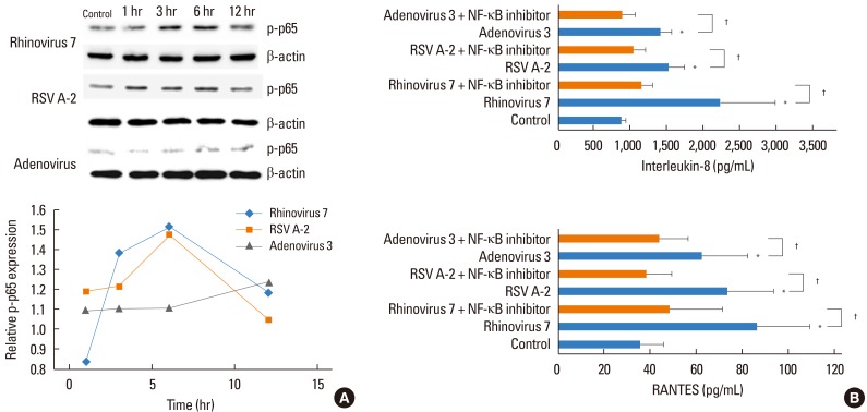
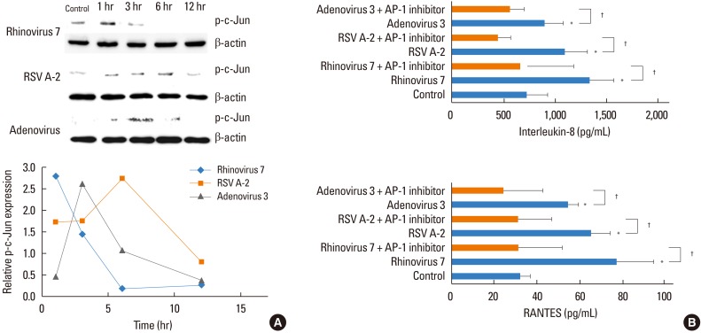
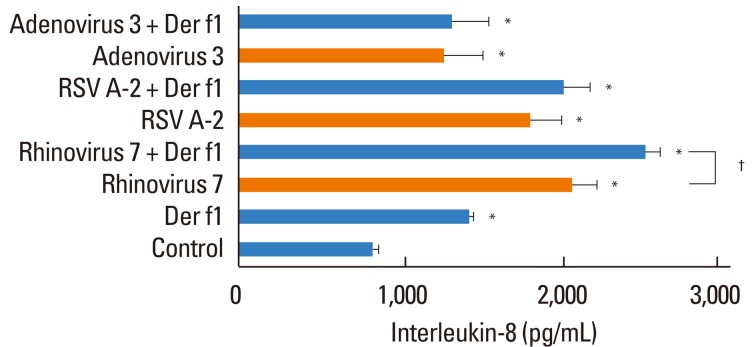
Methods: A549 human airway epithelial cells were infected with either rhinovirus serotype 7, respiratory syncytial virus (RSV)-A2 strain, or adenovirus serotype 3 and analyzed for interleukin (IL)-8 and regulated on activation, normal T-cell expressed and secreted (RANTES) release and mRNA expression. Additionally, activation of nuclear factor (NF)-κB and activator protein (AP)-1 were evaluated. The release of IL-8 and RANTES was also measured in cells stimulated simultaneously with a virus and the HDM allergen, Der f1.

Results: HRV caused greater IL-8 and RANTES release and mRNA expression compared with either RSV or adenovirus. NF-κB and AP-1 were activated in these processes. Cells incubated with a virus and Der f1 showed an increased IL-8 release. However, compared with cells incubated with virus alone as the stimulator, only HRV with Der f1 showed a statistically significant increase.

Conclusions: IL-8 and RANTES were induced to a greater extent by HRV compared with other viruses, and only HRV with Der f1 acted synergistically to induce bronchial epithelial IL-8 release. These findings may correspond with the fact that rhinoviruses are identified more frequently than other viruses in cases of acute exacerbation of asthma.

Keywords: Der f1; IL-8; Rhinovirus; asthma; regulated on activation, normal T-cell expressed and secreted.


Conflict of interest statement

There are no financial or other issues that might lead to conflict of interest.

Figures

Fig. 1
Fig. 1
(A) Effects of viral infection on IL-8 release (mean [standard deviation]) from A549 cells, as assessed by ELISA. The increase in IL-8 release was as follows: rhinovirus>respiratory syncytial virus (RSV)=adenovirus>control. A549 cells were infected at 102 50% tissue culture infectious dose (TCID50)/mL (n=3). *P<0.05, compared with the control, P<0.05, compared with adenovirus, P<0.05, compared with RSV. (B) IL-8 mRNA expression in A549 cells infected with rhinovirus, RSV, or adenovirus at 10-1 to 102 TCID50/mL. The mRNA expression of IL-8 occurred in decreasing order at 24 hr after infection with rhinovirus, RSV, and adenovirus. TNF α (10 ng/mL) and IL-4 (10 ng/mL) were stimulators for the positive control. Glyceraldehyde-3-phosphate dehydrogenase (GAPDH) was used as a housekeeping gene.
Fig. 2
Fig. 2
(A) Effects of viral infection on RANTES release (mean [standard deviation]) from A549 cells, as assessed by ELISA. The increase in RANTES release was as follows: rhinovirus>RSV>adenovirus=control. A549 cells were infected at 102 TCID50/mL (n=3). *P<0.05, compared with the control, P<0.05, compared with adenovirus, P<0.05, compared with RSV. (B) RANTES mRNA expression in A549 cells infected with rhinovirus, RSV, or adenovirus at 10-1 to 102 TCID50/mL. The mRNA expression of RANTES occurred in decreasing order at 24 hr after infection with rhinovirus, RSV, and adenovirus. TNF-α (10 ng/mL) and IL-4 (10 ng/mL) were stimulators for the positive control. GAPHD was used as a housekeeping gene.
Fig. 3
Fig. 3
(A) Phosphorylated subunits of NF-κB (pp65) were analyzed by SDS-PAGE. The band densities were determined by densitometry. Relative pp65 expressions were calculated as the pp65/β-actin ratio of control. (B) Effect of NF-κB inhibitor on the release of IL-8 and RANTES from A549 cells after 48 hr infection assessed by ELISA. All three viruses induced IL-8 and RANTES release compared with the control medium (*P<0.05). Fifty µM pyrollidine dithiocarbamate (PDTC) suppressed the release of IL-8 and RANTES in all three viruses compared with virus only (P<0.05). Data are from three independent experiments and results are presented as the mean±standard deviation.
Fig. 4
Fig. 4
(A) Phosphorylated subunits of AP-1 (p-c-Jun) were analyzed by SDS-PAGE. The band densities were determined by densitometry. Relative p-c-Jun expressions were calculated as the p-c-Jun/β-actin ratio of control. (B) Effect of AP-1 inhibitor on the release of IL-8 and RANTES from A549 cells 48 hr after infection assessed by ELISA. All three viruses induced increased IL-8 and RANTES release compared with the control medium (*P<0.05). Fifty µM SP600125C suppressed induction of IL-8 and RANTES by all three viruses compared with virus only (P<0.05). Data are from three independent experiments and results are presented as mean±standard deviation.
Fig. 5
Fig. 5
The release of IL-8 (mean [standard deviation]) from A549 cells after viral infection with or without exposure to dust mite allergen Der f1 (1 µg/mL) as assessed by ELISA. All stimuli induced an increase in IL-8 release compared with the control medium at 24 hr after infection. Only rhinovirus with Der f1 induced a greater increase in IL-8 release compared with single viral stimulation. Each virus was infected with 102 TCID50/mL (n=3). *P<0.05, compared with the control, P<0.05, virus alone versus in combination with Der f1.

References

    1. Kim WK, Gern JE. Updates in the relationship between human rhinovirus and asthma. Allergy Asthma Immunol Res. 2012;4:116–121. - PMC - PubMed
    1. Kotaniemi-Syrjänen A, Vainionpää R, Reijonen TM, Waris M, Korhonen K, Korppi M. Rhinovirus-induced wheezing in infancy--the first sign of childhood asthma? J Allergy Clin Immunol. 2003;111:66–71. - PMC - PubMed
    1. Jackson DJ, Johnston SL. The role of viruses in acute exacerbations of asthma. J Allergy Clin Immunol. 2010;125:1178–1187. quiz 1188-9. - PMC - PubMed
    1. Wark PA, Johnston SL, Moric I, Simpson JL, Hensley MJ, Gibson PG. Neutrophil degranulation and cell lysis is associated with clinical severity in virus-induced asthma. Eur Respir J. 2002;19:68–75. - PubMed
    1. Wenzel S. Mechanisms of severe asthma. Clin Exp Allergy. 2003;33:1622–1628. - PubMed