. 2014 Jan 20;20(3):391-402.
doi: 10.1089/ars.2012.4634. Epub 2013 Sep 5.

Keratinocyte growth factor and glucocorticoid induction of human peroxiredoxin 6 gene expression occur by independent mechanisms that are synergistic

Affiliations

Keratinocyte growth factor and glucocorticoid induction of human peroxiredoxin 6 gene expression occur by independent mechanisms that are synergistic

Ibrul Chowdhury et al. Antioxid Redox Signal. .

Abstract

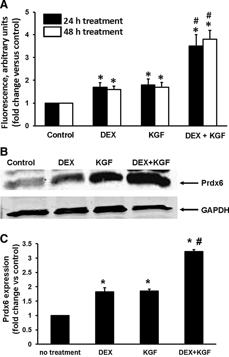
Aims: Peroxiredoxin 6 (Prdx6), a 1-cys Prdx has both peroxidase and phospholipase A2 activities, protecting against oxidative stress and regulating pulmonary surfactant phospholipid metabolism. This study determined the mechanism by which keratinocyte growth factor (KGF) and the glucocorticoid analogue, dexamethasone (Dex), induce increased Prdx6 expression.

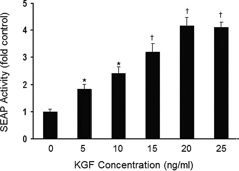
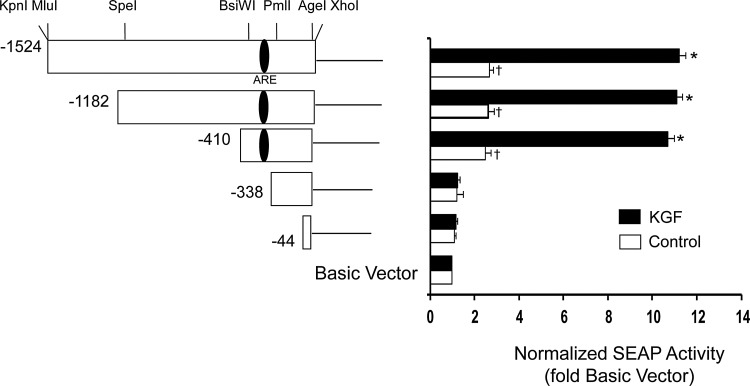
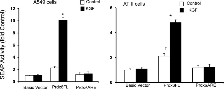
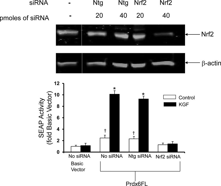
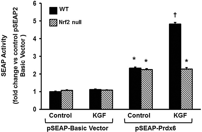
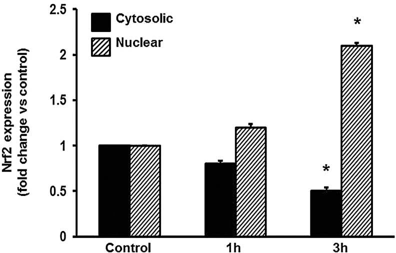
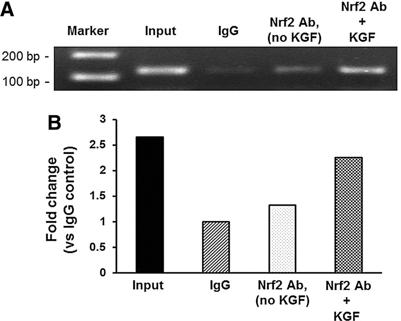
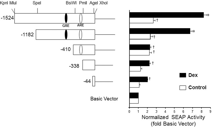
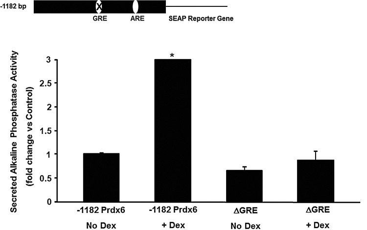
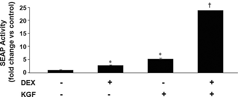
Results: Transcriptional activation by KGF in both A549 lung adenocarcinoma cells and rat lung alveolar epithelial type II (ATII) cells utilizes an antioxidant response element (ARE), located between 357 and 349 nucleotides before the PRDX6 translational start, that is also necessary for upregulation of the human PRDX6 promoter in response to oxidative stress. Activation is mediated by binding of the transcription factor, Nrf2, to the ARE as shown by experiments using siRNA against Nrf2 and by transfecting ATII cells isolated from lungs of Nrf2 null mice. KGF triggers the migration of Nrf2 from cytoplasm to nucleus where it binds to the PRDX6 promoter as shown by chromatin immunoprecipitation assays. Activation of transcription by Dex occurs through a glucocorticoid response element located about 750 nucleotides upstream of the PRDX6 translational start.

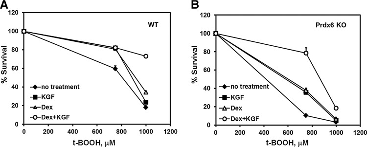
Innovation: This study demonstrates that KGF can activate an ARE in a promoter without reactive oxygen species involvement and that KGF and Dex can synergistically activate the PRDX6 promoter and protect cells from oxidative stress.

Conclusion: These two different activators work through different DNA elements. Their combined effect on transcription of the reporter gene is synergistic; however, at the protein level, the combined effect is additive and protects cells from oxidative damage.


Figures

<b>FIG. 1.</b>
FIG. 1.
PRDX6 promoter activity in A549 cells responds to keratinocyte growth factor (KGF) in a dose-dependent manner. A549 cells, transiently transfected with pSEAP2 reporter plasmid containing the 1.5 kb peroxiredoxin 6 (PRDX6) upstream region, were cultured for 24 h, then treated with a range of KGF concentrations (5 ng/ml-25 ng/ml). Secreted alkaline phosphatase (SEAP) activity was determined 24 h later. The values shown are the mean±SE (n=3). *p<0.05, †p<0.01, compared with the control (untreated) cells.
<b>FIG. 2.</b>
FIG. 2.
Basal and KGF-inducible activity of PRDX6 promoter deletions. Left panel, a series of constructs were generated by restriction enzyme digestion of the 1.5 kb DNA fragment derived from the 5′-flanking region of PRDX6 in the promoterless pSEAP2-Basic vector. Numbers marking the 5′ ends of the deletions are relative to the translational start. The approximate location of the putative antioxidant response element (ARE) is shown within the promoter. Right panel, A549 cells were transiently transfected with reporter vectors and then incubated for 24 h, followed by 20 ng/ml of KGF treatment (or no treatment in control samples). SEAP activity was determined 24 h later. Mean±SE (n=3) are shown. *p<0.01 compared with vehicle control, †p<0.05 compared with pSEAP2-basic vector (no insert).
<b>FIG. 3.</b>
FIG. 3.
Role of the ARE in PRDX6 gene expression. Deletion of the ARE was generated by site-directed mutagenesis of the full-length PRDX6 promoter in the pSEAP2-Basic vector. Cells were transiently transfected with reporter vectors; after 24 h, cells were incubated with 20 ng/ml of KGF. SEAP activity was determined 24 h after KGF treatment. Cells were then harvested, and reporter gene assays performed. Prdx6FL, full-length PRDX6 promoter (1.5 kb); Prdx6ΔARE, full-length PRDX6 promoter with ARE deletion. Transfection and treatment of A549 cells (left panel) or rat primary alveolar type II (ATII) cells (right panel) with 20 ng/ml KGF for 24 h. Values shown are mean±SE (n=3) with untreated pSEAP2-basic vector set to 1. †p<0.05 *p<0.01 compared with untreated control.
<b>FIG. 4.</b>
FIG. 4.
siRNA knockdown of Nrf2 in A549 cells blocks induction of Prdx6 expression by KGF. Upper panel, targeted siRNA knockdown reduces Nrf2 in A549 cells. A549 cells were transfected with 20 or 40 pmoles of non-targeting siRNA (Ntg) or siRNA directed against Nrf2 (Nrf2) and cultured for 72 h. β-actin was used as a loading control. The gel shown was edited to excise irrelevant lanes. The vertical gray line marks the location of the excised portion. Lower panel, A549 cells were transfected with the pSEAP2-Basic plasmid (Basic vector) or the same plasmid with the full-length PRDX6 promoter (Prdx6FL) and, where indicated, siRNA; Ntg as a negative control or siRNA targeted against Nrf2. Assays were performed 24 h after treatment. Control; cells not treated with KGF. KGF; cells treated with 20 ng/ml KGF. Values shown are mean±SE (n=3) with untreated pSEAP2-Basic vector set to 1. *p<0.05 compared with vehicle control, †p<0.05 compared with pSEAP2-Basic vector.
<b>FIG. 5.</b>
FIG. 5.
KGF treatment does not stimulate promoter activity in Nrf2 knockout mice. Cells were transiently transfected with the indicated reporter vectors; after 24 h, cells were incubated with 20 ng/ml of KGF. SEAP activity was determined 24 h after treatment. Cells were then harvested, reporter gene assays performed and transfection efficiency was normalized to cotransfected β-galactosidase. The values are the mean±SE (n=3).*p<0.05, †p<0.01, compared with pSEAP-2-basic vector in the nontreated cells.
<b>FIG. 6.</b>
FIG. 6.
KGF-induced Nrf2 nuclear translocation in A549 cells. The content of Nrf2 cytosolic and nuclear fractions was determined in the indicated subcellular fraction by western blot analysis. Cells were exposed to 20 ng/ml KGF for the indicated times. Values shown are percent of the values in untreated cells. GAPDH (cytosolic fraction) and PCNA (nuclear fraction) were used as loading controls. Mean±SE (n=3). *p<0.05 compared with control.
<b>FIG. 7.</b>
FIG. 7.
Chromatin immunoprecipitation (ChIP) assay of the PRDX6 promoter ARE in response to KGF treatment. (A) A549 cells treated with 20 ng/ml KGF for 24 h were processed for ChIP assays using the primer pairs described in Materials and Methods section. Lane 1: Marker; 100 bp ladder: Input; results obtained from DNA that was polymerase chain reaction (PCR) amplified from chromatin extracts before immunoprecipitation, IgG; PCR results obtained after immunoprecipitation with rabbit immunoglobulin G. Nrf2 AB, no KGF; PCR results obtained after immunoprecipitation with Nrf2 antibody; in unstimulated cells: Nrf2 Ab+KGF; PCR results obtained after immunoprecipitation with Nrf2 antibody in cells treated with KGF. Results are representative for n=3. (B) Densitometric quantification of ChIP assay. Quantification of bands in A was performed using the ImageJ built-in measure function. Values are presented as a fold change versus IgG control. Data are shown as fold change versus IgG control.
<b>FIG. 8.</b>
FIG. 8.
Basal and dexamethasone (Dex)-inducible activity of PRDX6 promoter deletions. Left panel, a series of constructs were generated by restriction enzyme digestion of the 1.5 kb DNA fragment derived from the 5′-flanking region of PRDX6 in the promoterless pSEAP2-Basic vector. Numbers marking the 5′ ends of the deletions are relative to the translational start. The approximate location of the putative glucocorticoid response element (GRE) (and ARE) are shown within the promoter. Right panel, A549 cells were transiently transfected with reporter vectors and then incubated for 24 h, followed by 1 μM Dex treatment (or no treatment in control samples). SEAP activity was determined 24 h later. Mean±SE (n=3) are shown. *p<0.01 compared with vehicle control, †p<0.05 compared with pSEAP2-basic vector (no insert).
<b>FIG. 9.</b>
FIG. 9.
Role of the GRE in PRDX6 gene expression. Deletion of the GRE was generated by site-directed mutagenesis of the 1.2 kb human PRDX6 promoter in the pSEAP2-Basic vector. Cells were transiently transfected with reporter vectors; after 24 h, cells were incubated with1 μM Dex. SEAP activity was determined 24 h after Dex treatment. Cells were then harvested, and reporter gene assays performed. −1182 Prdx, undeleted 1.2 kb PRDX6 promoter; ΔGRE, 1.2 kb PRDX6 promoter with GRE deletion. Values shown are mean±SE (n=3) with untreated pSEAP2-Basic vector (control) set to 1. *p<0.05 compared with pSEAP2-basic vector.
<b>FIG. 10.</b>
FIG. 10.
Synergistic effect of KGF and Dex on induction of PRDX6 promoter activity. The effect of the combination of KGF and Dex on reporter gene activity in response to the human PRDX6 promoter is shown. The effect of each individual treatment is shown for comparison. The basal level of reporter gene activity is set to 1. Cells were transiently transfected with reporter vectors; after 24 h, cells were incubated with 20 ng/ml of KGF, 1 μM Dex, or both. SEAP activity was determined 24 h after treatment. Cells were then harvested, and reporter gene assays performed. Mean±SE (n=3) are shown. *p<0.05 compared with untreated control, †p<0.01 compared with untreated control.
<b>FIG. 11.</b>
FIG. 11.
Upregulation of Prdx6 protein expression in A549 cells under the Dex and KGF treatments. (A) Determined by flow cytometry (FCM). Geometric mean fluorescence (arbitrary units) of Prdx6 intracellular content in control (untreated) versus Dex or KGF-treated A549 cells by FCM analysis. Cells underwent KGF (20 ng/ml), Dex (10−6 M), or combined Dex+KGF treatment for 24 or 48 h. Geometric Mean fluorescence of Prdx6 intracellular content was normalized by 50,000 events. Bars demonstrate the fold increase in Prdx6 intracellular concentration in KGF/Dex-treated A549 cells in comparison with basic fluorescence of untreated cells, shown as 1. Mean±SE (n=3) *p<0.05 compared with untreated control; #p<0.001 compared with a single treatment. (B) Western blot analysis of Prdx6 expression in A549 cells after 48 h treatment with 20 ng/ml KGF, 1 μM Dex, or combined treatment. (C) Quantification of western blot analysis of Prdx6 expression in KGF and/or Dex-treated A549 cells. For each sample, Prdx6 expression was normalized for the expression of β-actin or GAPDH. The amount of Prdx6 in non-treated (control) A549 cells is shown as 1. Mean±SE (n=3) *p<0.001 versus control; #p<0.001 versus single treatment.
<b>FIG. 12.</b>
FIG. 12.
The effect of KGF and Dex on wild-type (WT) and Prdx6 null mouse ATII cell survival from oxidative stress. Comparison of survival rates for WT (A) or Prdx6 null (B) cells after treatment with indicated concentrations of tBOOH for 24 h were measured by the neutral red assay. Where indicated, cells received a 24 h pretreatment with 20 ng/ml KGF and/or 1 μM Dex. Data shown are mean±SE (n=5). *p<0.0001 compared with other points at the same concentration of tBOOH.

References

    1. Bao S, Wang Y, Sweeney P, Chaudhuri A, Doseff AI, Marsh CB, and Knoell DL. Keratinocyte growth factor induces Akt kinase activity and inhibits Fas-mediated apoptosis in A549 lung epithelial cells. Am J Physiol Lung Cell Mol Physiol 288: L36–L42, 2005 - PubMed
    1. Barazzone C, Donati YR, Rochat AF, Vesin C, Kan CD, Pache JC, and Piguet PF. Keratinocyte growth factor protects alveolar epithelium and endothelium from oxygen-induced injury in mice. Am J Pathol 154: 1479–1487, 1999 - PMC - PubMed
    1. Bortnick AE, Favari E, Tao JQ, Francone OL, Reilly M, Zhang Y, Rothblat GH, and Bates SR. Identification and characterization of rodent ABCA1 in isolated type II pneumocytes. Am J Physiol Lung Cell Mol Physiol 285: L869–L878, 2003 - PubMed
    1. Braun S, Hanselmann C, Gassmann MG, auf dem Keller U, Born-Berclaz C, Chan K, Kan YW, and Werner S. Nrf2 transcription factor, a novel target of keratinocyte growth factor action which regulates gene expression and inflammation in the healing skin wound. Mol Cell Biol 22: 5492–5505, 2002 - PMC - PubMed
    1. Chen JW, Dodia C, Feinstein SI, Jain MK, and Fisher AB. 1-Cys peroxiredoxin, a bifunctional enzyme with glutathione peroxidase and phospholipase A2 activities. J Biol Chem 275: 28421–28427, 2000 - PubMed

Publication types

MeSH terms

LinkOut - more resources