. 2013 Jul 1:14:31.
doi: 10.1186/1471-2121-14-31.

Chronic glucolipotoxic conditions in pancreatic islets impair insulin secretion due to dysregulated calcium dynamics, glucose responsiveness and mitochondrial activity

Affiliations

Chronic glucolipotoxic conditions in pancreatic islets impair insulin secretion due to dysregulated calcium dynamics, glucose responsiveness and mitochondrial activity

Baggavalli P Somesh et al. BMC Cell Biol. .

Abstract

Background: In the progression towards diabetes, glucolipotoxicity is one of the main causes of pancreatic beta cell pathology. The aim of this study was to examine the in vitro effects of chronic glucolipotoxic conditions on cellular responses in pancreatic islets, including glucose and fat metabolism, Calcium mobilization, insulin secretion and insulin content.

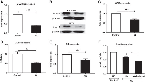
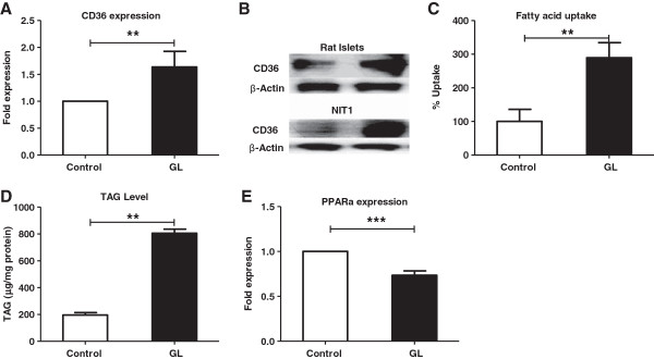
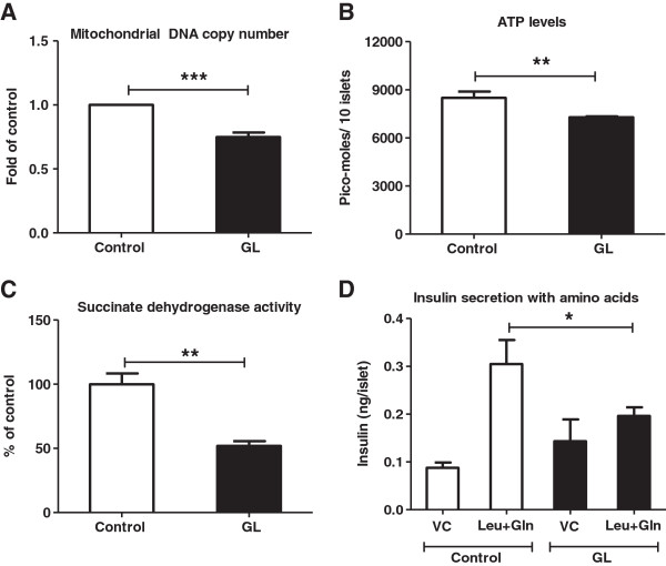
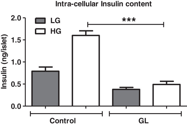
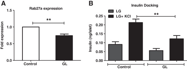
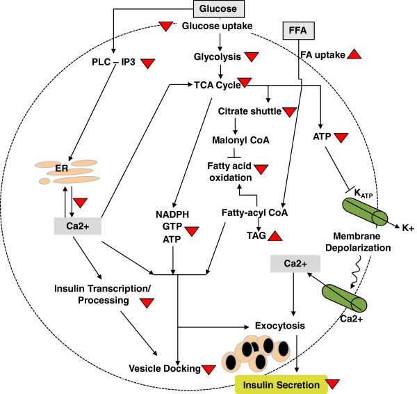
Results: Exposure of islets to chronic glucolipotoxic conditions decreased glucose stimulated insulin secretion in vitro. Reduced protein levels of Glut2/slc2a2, and decreased glucokinase and pyruvate carboxylase mRNA levels indicated a significant lowering in glucose sensing. Concomitantly, both fatty acid uptake and triglyceride accumulation increased significantly while fatty acid oxidation decreased. This general suppression in glucose metabolism correlated well with a decrease in mitochondrial number and activity, reduction in cellular ATP content and dampening of the TCA cycle. Further, we also observed a decrease in IP3 levels and lower Calcium mobilization in response to glucose. Importantly, chronic glucolipotoxic conditions in vitro decreased insulin gene expression, insulin content, insulin granule docking (to the plasma membrane) and insulin secretion.

Conclusions: Our results present an integrated view of the effects of chronic glucolipotoxic conditions on known and novel signaling events, in vitro, that results in reduced glucose responsiveness and insulin secretion.


Figures

Figure 1
Figure 1
Chronic glucolipotoxic conditions in vitro reduce GSIS. Rat islets were cultured under normal conditions (control) or with 16.7 mM glucose and 500 μM palmitate for 72 h (GL). Post 72 h, islets were treated with 2 mM glucose (LG) or 11 mM glucose (HG) for 2 h and secreted insulin was measured. Data are expressed as mean±SEM and statistical analysis was performed using the unpaired Student’s t-test. (*P<0.05, **P<0.01 and ***P<0.001, n=4).
Figure 2
Figure 2
In vitro chronic glucolipotoxicity impairs glucose uptake/metabolism. Rat islets or NIT1 cells were cultured under normal conditions (control) or with 16.7 mM glucose and 500 μM palmitate for 72 h (GL). Post 72 h, RNA was isolated from islets and the mRNA levels of Glut2/slc2a2 (A), GCK (C) and PC (E) were measured as described in the materials and methods. Protein levels of Glut2/slc2a2 (B) were measured in both islets and NIT-1 cells by western blotting using anti-Glut2/slc2a2 antibodies. Glucose uptake (D) was measured NIT-1 cells using 2-NBDG, a non-metabolized fluorescent analog as described in the materials and methods. (F) For insulin secretion in the presence of ATP citrate lyase (ACLY) inhibitor (Radicicol), post 72 h treatment islets were treated with 11 mM glucose (HG) with/without 50 μM inhibitor for 2 h and secreted insulin were measured. Data are expressed as mean±SEM and statistical analysis was performed using the unpaired Student’s t-test. (*P<0.05, **P<0.01 and ***P<0.001, n=4).
Figure 3
Figure 3
Chronic glucolipotoxic conditions in vitro impair fatty acid uptake/ metabolism. Rat islets and NIT1 cells were cultured under normal conditions (control) or with 16.7 mM glucose and 500 μM palmitate for 72 h (GL). Post 72 h, RNA was isolated from islets and the mRNA levels of cd36 (A) and PPARα (E) were measured as described in the materials and methods. Protein levels of CD36 in both islets and NIT-1 cells (B) were measured by western blotting using anti-CD36 antibodies. Fatty acid uptake (C) was measured in NIT-1 cells using green-fluorescent BODIPY dyes, a non-metabolized fluorescent labelled fatty acid analog as described in the materials and methods. For triglyceride estimation, post incubation cells were lysed and cellular triglyceride levels were estimated and normalized to total protein content (D). Data are expressed as mean±SEM and statistical analysis was performed using the unpaired Student’s t-test. (*P<0.05, **P<0.01 and ***P<0.001, n=4).
Figure 4
Figure 4
Chronic glucolipotoxicity in vitro reduces mitochondrial number and activity. Rat islets and NIT1 cells were cultured under normal conditions (control) or with 16.7 mM glucose and 500 μM palmitate for 72 h (GL). Post 72 h, total DNA was isolated from islets and mitochondrial DNA copy number (A) was estimated by assessing mtCox1 (mitochondrial gene) copy number normalized to HPRT (nuclear gene). Post treatment, islet ATP levels (B) were estimated after 60min induction with 11 mM (HG) as described in the materials and methods. Both SDH activity in NIT-1 cells (C) and amino acids (5 mM of leucine and 5 mM of glutamine) mediated insulin secretion (D) in islets were measured as described in the materials and methods. VC is vehicle control with 2 mM glucose and without amino acids. Data are expressed as mean±SEM and statistical analysis was performed using the unpaired Student’s t-test. (*P<0.05, **P<0.01 and ***P<0.001, n=4).
Figure 5
Figure 5
Chronic glucolipotoxicity in vitro impairs calcium release. Rat islets and NIT-1 cells were cultured under normal conditions (control) or with 16.7 mM glucose and 500 μM palmitate for 72 h (GL). Post 72 h, islet IP3 levels (A) were estimated in the presence of 11 mM (HG) as described in the materials and methods. For cytosolic calcium mobilization assay (B and C), post treatment NIT-1 cells were loaded with Fluo-3AM calcium indicator dye for 1 h. After washing the cells, they were treated with either low (2.5 mM) or high (16.7 mM) glucose and the change in fluorescence was estimated as described in the materials and methods. Data are expressed as mean±SEM and statistical analysis was performed using the unpaired Student’s t-test. (*P<0.05, **P<0.01 and ***P<0.001, n=4).
Figure 6
Figure 6
Chronic glucolipotoxic conditions in vitro reduce intracellular insulin content. Rat islets were cultured under normal conditions (control) or with 16.7mM glucose and 500 μM palmitate for 72 h (GL). Post 72 h, islets were treated with 2 mM glucose (LG) or 11 mM glucose (HG) for 2 h. Islets were washed, lysed and islet insulin content was measured. Data is expressed as mean±SEM and statistical analysis was performed using the unpaired Student’s t-test. (*P<0.05, **P<0.01 and ***P<0.001, n=4).
Figure 7
Figure 7
In vitro chronic glucolipotoxicity reduces insulin docking and exocytosis. Rat islets were cultured under normal conditions (control) or with 16.7 mM glucose and 500 μM palmitate for 72 h (GL). Post 72 h, RNA was isolated from islets and the mRNA levels of Rab27a (A) were measured as described in the materials and methods. For measurement of insulin granule docking (B) following incubation under chronic glucolipotoxic conditions, islets were treated with LG (2.5 mM glucose) with or without 30 mM KCl for 30 min. Secreted insulin was measured as described in the materials and methods. Data are expressed as mean±SEM and statistical analysis was performed using the unpaired Student’s t-test. (*P<0.05, **P<0.01 and ***P<0.001, n=4).
Figure 8
Figure 8
Model for impact of in vitro chronic glucolipotoxicity on cellular processes in pancreatic beta cells. Model describes the signaling pathways involved in glucose and fat metabolism with focus on their coupling to insulin secretion. We found that under chronic glucolipotoxic conditions in vitro, multiple signaling events involved in glucose and fat metabolism are dysregulated leading to impaired insulin synthesis, content and secretion. The effects of chronic glucolipotoxic conditions on the signaling events involved are shown using upward arrowheads (representing an increase) and downward arrowheads (representing a decrease). In the model, arrows indicate activation and blunt arrows indicate inhibition.

References

    1. Prentki M, Nolan CJ. Islet beta cell failure in type 2 diabetes. J Clin Invest. 2006;116:1802–1812. doi: 10.1172/JCI29103. - DOI - PMC - PubMed
    1. Defronzo RA. Banting Lecture. From the triumvirate to the ominous octet: a new paradigm for the treatment of type 2 diabetes mellitus. Diabetes. 2009;58:773–795. doi: 10.2337/db09-9028. - DOI - PMC - PubMed
    1. Poitout V, Amyot J, Semache M, Zarrouki B, Hagman D, Fontés G. Glucolipotoxicity of the pancreatic beta cell. Biochim Biophys Acta. 1801;2010:289–298. - PMC - PubMed
    1. Poitout V, Robertson RP. Secondary beta-cell failure in type 2 diabetes-a convergence of glucotoxicity and lipotoxicity. Endocrinology. 2002;143:339–342. doi: 10.1210/en.143.2.339. - DOI - PubMed
    1. El-Assaad W, Buteau J, Peyot ML, Nolan C, Roduit R, Hardy S, Joly E, Dbaibo G, Rosenberg L, Prentki M. Saturated fatty acids synergize with elevated glucose to cause pancreatic beta-cell death. Endocrinol. 2003;144:4154–4163. doi: 10.1210/en.2003-0410. - DOI - PubMed

Publication types

MeSH terms