

Genomic distribution of SINEs in Entamoeba histolytica strains: implication for genotyping
- PMID: 23815468
- PMCID: PMC3716655
- DOI: 10.1186/1471-2164-14-432
Genomic distribution of SINEs in Entamoeba histolytica strains: implication for genotyping
Abstract
Background: The major clinical manifestations of Entamoeba histolytica infection include amebic colitis and liver abscess. However the majority of infections remain asymptomatic. Earlier reports have shown that some E. histolytica isolates are more virulent than others, suggesting that virulence may be linked to genotype. Here we have looked at the genomic distribution of the retrotransposable short interspersed nuclear elements EhSINE1 and EhSINE2. Due to their mobile nature, some EhSINE copies may occupy different genomic locations among isolates of E. histolytica possibly affecting adjacent gene expression; this variability in location can be exploited to differentiate strains.
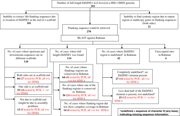
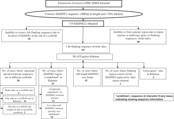
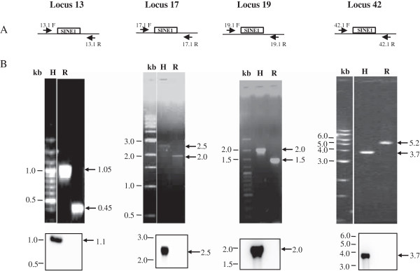
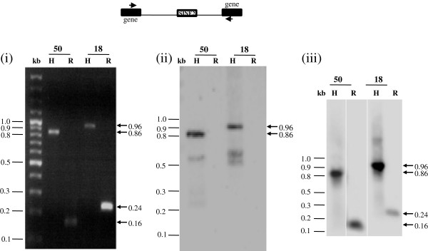
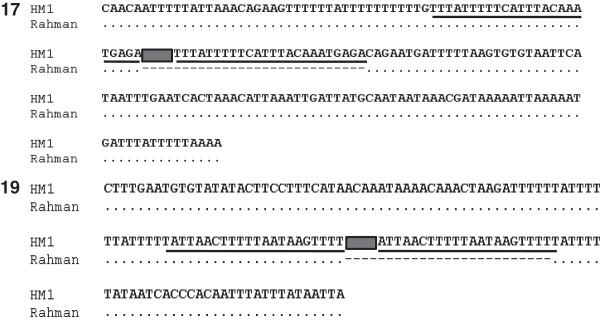
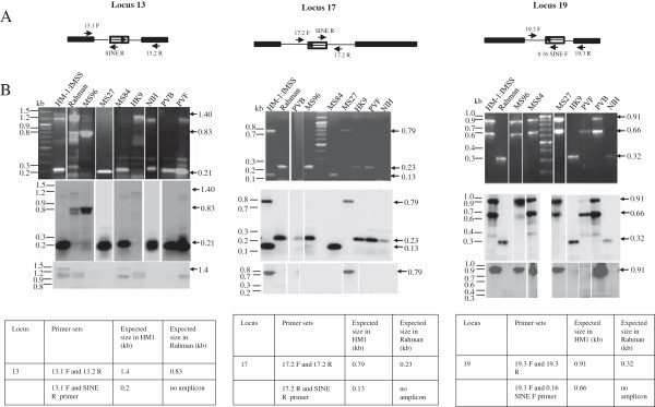
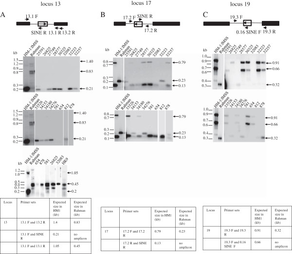
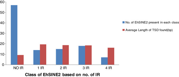
Results: We have looked for EhSINE1- and EhSINE2-occupied loci in the genome sequence of Entamoeba histolytica HM-1:IMSS and searched for homologous loci in other strains to determine the insertion status of these elements. A total of 393 EhSINE1 and 119 EhSINE2 loci were analyzed in the available sequenced strains (Rahman, DS4-868, HM1:CA, KU48, KU50, KU27 and MS96-3382. Seventeen loci (13 EhSINE1 and 4 EhSINE2) were identified where a EhSINE1/EhSINE2 sequence was missing from the corresponding locus of other strains. Most of these loci were unoccupied in more than one strain. Some of the loci were analyzed experimentally for SINE occupancy using DNA from strain Rahman. These data helped to correctly assemble the nucleotide sequence at three loci in Rahman. SINE occupancy was also checked at these three loci in 7 other axenically cultivated E. histolytica strains and 16 clinical isolates. Each locus gave a single, specific amplicon with the primer sets used, making this a suitable method for strain typing. Based on presence/absence of SINE and amplification with locus-specific primers, the 23 strains could be divided into eleven genotypes. The results obtained by our method correlated with the data from other typing methods. We also report a bioinformatic analysis of EhSINE2 copies.
Conclusions: Our results reveal several loci with extensive polymorphism of SINE occupancy among different strains of E. histolytica and prove the principle that the genomic distribution of SINEs is a valid method for typing of E. histolytica strains.
Figures
Similar articles
-
SINE polymorphism reveals distinct strains of Entamoeba histolytica from North India.Exp Parasitol. 2017 Apr;175:28-35. doi: 10.1016/j.exppara.2017.01.007. Epub 2017 Jan 26. Exp Parasitol. 2017. PMID: 28131659
-
Differential distribution of a SINE element in the Entamoeba histolytica and Entamoeba dispar genomes: role of the LINE-encoded endonuclease.BMC Genomics. 2011 May 25;12:267. doi: 10.1186/1471-2164-12-267. BMC Genomics. 2011. PMID: 21612594 Free PMC article.
-
Identification of differentially expressed genes in virulent and nonvirulent Entamoeba species: potential implications for amebic pathogenesis.Infect Immun. 2006 Jan;74(1):340-51. doi: 10.1128/IAI.74.1.340-351.2006. Infect Immun. 2006. PMID: 16368989 Free PMC article.
-
The non-LTR retrotransposons of Entamoeba histolytica: genomic organization and biology.Mol Genet Genomics. 2022 Jan;297(1):1-18. doi: 10.1007/s00438-021-01843-5. Epub 2022 Jan 9. Mol Genet Genomics. 2022. PMID: 34999963 Review.
-
Molecular methods for diagnosis of Entamoeba histolytica in a clinical setting: an overview.Exp Parasitol. 2007 May;116(1):35-43. doi: 10.1016/j.exppara.2006.11.005. Epub 2006 Dec 26. Exp Parasitol. 2007. PMID: 17189632 Free PMC article. Review.
Cited by
-
Pathogenicity and virulence of Entamoeba histolytica, the agent of amoebiasis.Virulence. 2023 Dec;14(1):2158656. doi: 10.1080/21505594.2022.2158656. Virulence. 2023. PMID: 36519347 Free PMC article. Review.
-
Evolutionary genomics and population structure of Entamoeba histolytica.Comput Struct Biotechnol J. 2014 Oct 31;12(20-21):26-33. doi: 10.1016/j.csbj.2014.10.001. eCollection 2014 Nov. Comput Struct Biotechnol J. 2014. PMID: 25505504 Free PMC article. Review.
-
Attenuation of In Vitro and In Vivo Virulence Is Associated with Repression of Gene Expression of AIG1 Gene in Entamoeba histolytica.Pathogens. 2023 Mar 21;12(3):489. doi: 10.3390/pathogens12030489. Pathogens. 2023. PMID: 36986411 Free PMC article.
-
Amoebiasis: Advances in Diagnosis, Treatment, Immunology Features and the Interaction with the Intestinal Ecosystem.Int J Mol Sci. 2023 Jul 21;24(14):11755. doi: 10.3390/ijms241411755. Int J Mol Sci. 2023. PMID: 37511519 Free PMC article. Review.
References
-
- World Health Organization. Amoebiasis. Wkly Epidemiol Rec. 1997;72:97–100. - PubMed
Publication types
MeSH terms
Substances
LinkOut - more resources
Full Text Sources
Other Literature Sources
