

Precision of posttraumatic primary orbital reconstruction using individually bent titanium mesh with and without navigation: a retrospective study
- PMID: 23815979
- PMCID: PMC3750456
- DOI: 10.1186/1746-160X-9-18
Precision of posttraumatic primary orbital reconstruction using individually bent titanium mesh with and without navigation: a retrospective study
Abstract
Background: The aim of orbital wall reconstruction is to reestablish anatomically exact orbital volumes to avoid long-term complications. Navigation could facilitate complex reconstructions.
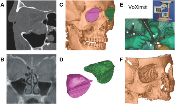
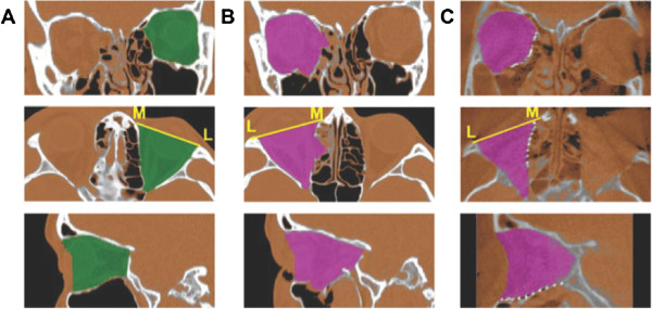
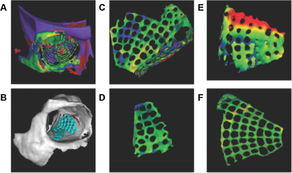
Methods: Quality of the orbital reconstruction (n=94) was measured based on (A) volume changes and (B) on 3D shape deviations compared to the unaffected side. Volume analysis included segmentation of the orbital cavity in the pre- and post-operative 3D data set (VoXim®, IVS Solutions, Germany), and shape analysis was performed by vector-based 3D tools (Comparison®, 3Dshape, Germany).
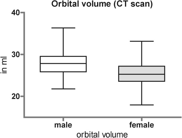
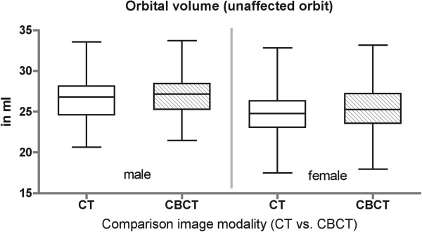
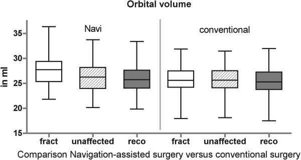
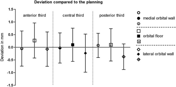
Results: Orbital volume of the unaffected side ranged from 26.6 ml±2.8 ml in male and 25.2 ml±2.6 ml in female (CT). Significant orbital enlargement was found in orbital fractures with involvement of the posterior third of the orbital floor and in comminuted fracture pattern. Reconstructed orbital volume ranged from 26.9±2.7 ml in male and 24.26±2.5 ml in female (CBCT). 3D Analysis of the color mapping showed minor deviations compared to the mirrored unaffected side.
Conclusion: Measurements demonstrate that even in comminuted orbital fractures true-to-original reconstruction is feasible.
Figures
References
-
- Kolk A, Pautke C, Schott V, Ventrella E, Wiener E, Ploder O. et al. Secondary post-traumatic enophthalmos: high-resolution magnetic resonance imaging compared with multislice computed tomography in postoperative orbital volume measurement. J Oral Maxillofac Surg. 2007;65(10):1926–1934. doi: 10.1016/j.joms.2006.06.269. - DOI - PubMed
-
- Dolynchuk KN, Tadjalli HE, Manson PN. Orbital volumetric analysis: clinical application in orbitozygomatic complex injuries. J Craniomaxillofac Trauma. 1996;2(2):56–63. discussion 64. - PubMed
Publication types
MeSH terms
Substances
LinkOut - more resources
Full Text Sources
Other Literature Sources
Medical
