. 2013 Jul 1:11:29.
doi: 10.1186/1477-5956-11-29. eCollection 2013.

Proteomic analyses of age related changes in A.BY/SnJ mouse hearts

Affiliations

Proteomic analyses of age related changes in A.BY/SnJ mouse hearts

Krishnatej Nishtala et al. Proteome Sci. .

Abstract

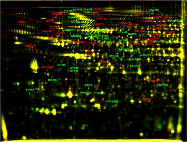
Background: A.BY/SnJ mice are used to study pathological alterations in the heart due to enteroviral infections. Since age is a well-known factor influencing the susceptibility of mice to infection, response to stress and manifestation of cardiovascular diseases, the myocardial proteome of A.BY/SnJ mice aged 1 and 4 months was comparatively studied using two dimensional-differential in-gel electrophoresis (2D-DIGE) and liquid chromatography tandem mass spectrometry (LC-MS/MS).

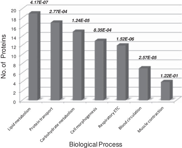
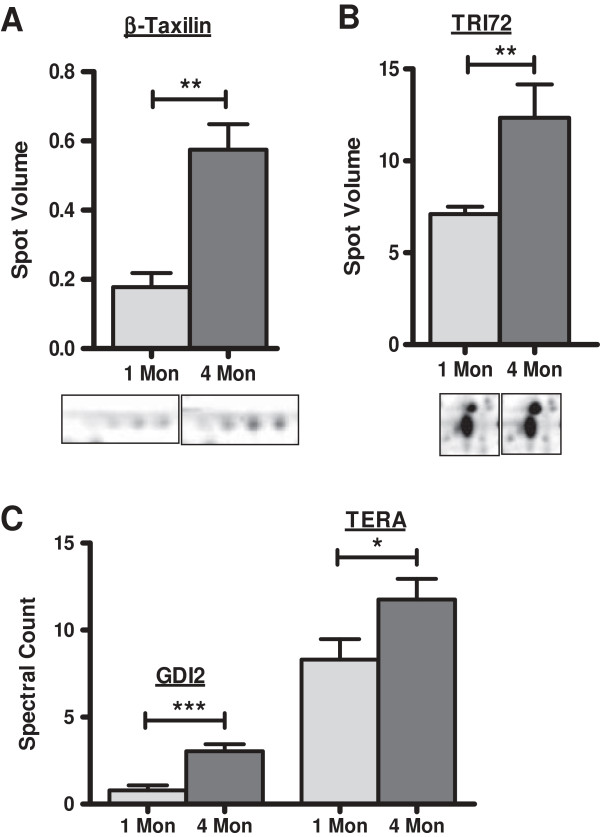
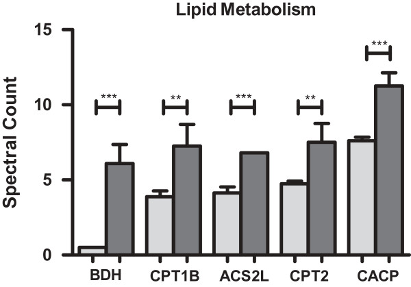
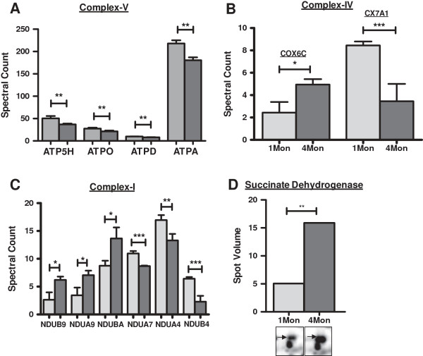
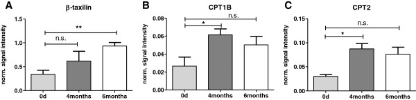
Results: Complementary analyses by 2D-DIGE and gel-free LC-MS/MS revealed 96 distinct proteins displaying age associated alterations in their levels. Proteins related to protein transport, and transport chain, lipid metabolism and fatty acid transport showed significant changes in 4 months old mouse hearts compared to juvenile hearts. Proteins involved in lipid metabolism and transport were identified at significantly higher levels in older mice and dysregulation of proteins of the respiratory transport chain were observed.

Conclusion: The current proteomics study discloses age dependent changes occurring in the hearts already in young mice of the strain A.BY/SnJ. Besides alterations in protein transport, we provide evidence that a decrease of ATP synthase in murine hearts starts already in the first months of life, leading to well-known low expression levels manifested in old mice thereby raising the possibility of reduced energy supply. In the first few months of murine life this seems to be compensated by an increased lipid metabolism. The functional alterations described should be considered during experimental setups in disease related studies.

Keywords: Aging; Differential in-gel electrophoresis; Hearts; LC-MS based quantitation; Murine model.


Figures

Figure 1
Figure 1
Changes in the protein profile of mouse hearts due to aging. Representative dual channel overlay of mice hearts 1 month old and 4 months old in the pH range of 4–7. A total of 1683 spots were detected on the gel using Delta 2D software (Decodon). Statistical analysis with Genespring (Agilent) revealed 176 spots with significantly altered intensities (p ≤ 0.05) of which 86 spots were identified by mass spectrometry. 40 (58) spots labeled in red showed ≥ 1.5 fold higher intensities, whereas 46 (118) spots labeled in green displayed > 1.5 fold lower intensities in the 4 months old mouse hearts compared to 1 month controls. Proteins which did not change in intensity appear in yellow.
Figure 2
Figure 2
Biological processes affected during aging in 4 months old mice heart in comparison to 1 month old animals. Results from gel-free LC-MS/MS and 2-D DIGE analysis were analyzed together for enrichment of biological processes using PANTHER. The most significantly affected pathways are represented in the graph in the order of number of proteins classified per protein class. The corresponding p-values were represented at the top of each column.
Figure 3
Figure 3
Increase in intracellular transport proteins identified by DIGE and gel-free LC-MS/MS analysis. Proteins involved in intracellular transport were identified in higher amounts in 4 months old mouse hearts compared to 1 month old hearts. Gray bars: 1 Month mice hearts, dark bars: 4 months mice hearts. A- Protein ß-taxilin (TXLNB) was identified in multiple spots and in higher amounts in 4 months hearts compared to control mice. B- Tripartite motif-72 (TRI72) showed approx. 1.7 fold increase in aging mice hearts. C- Proteins Rab GDP dissocation inhibitor 2 (GDI2) and Transitional endoplasmic reticulum ATPase (TERA) were identified in increasing amounts in aging mice hearts by gel-free LC-MS/MS analysis. * indicates p ≤ 0.1 ** indicates p ≤ 0.05 and *** indicates p ≤ 0.01.
Figure 4
Figure 4
Changes in lipid metabolism proteins in aging hearts by gel-free LC-MS/MS analysis based on spectral counts. High levels of proteins involved in lipid metabolism were observed in hearts of 4 months old mice in comparison to those of 1 month old animals suggesting increased energy production by lipid metabolism. Protein abundance in hearts of 1 month old mice: (grey bars), of 4 months old mice (dark bars). BDH-D-beta-hydroxybutyrate dehydrogenase, mitochondrial, CPT1B-carnitine O-palmitoyltransferase 1, muscle isoform, ACS2L-acetyl-coenzyme A synthetase 2-like, mitochondrial, CPT2-carnitine O-palmitoyltransferase 2, mitochondrial, CACP-carnitine O-acetyltransferase. ** indicates p ≤ 0.05 and *** indicates p ≤ 0.01.
Figure 5
Figure 5
Age dependent changes in abundance of proteins of the electron transport chain in mouse hearts. Different subunits of respiratory electron transport chain were affected due to aging in mice hearts. Grey bars: level in hearts of 1 month old animals, dark bars: level in hearts of 4 months old mice. A- subunits of ATP synthase (complex V) identified by gel-free analysis - ATP5H- ATP synthase subunit d, mitochondrial, ATPO- ATP synthase subunit O, mitochondrial, ATPD- ATP synthase subunit delta, mitochondrial, ATPA- ATP synthase subunit alpha, mitochondrial. B- cytochrome c oxidase subunit 6C (COX6C) and polypeptide 7A1, mitochondrial (CX7A1). C- subunits of complex I: NADH dehydrogenase [ubiquinone] 1 beta subcomplex subunit 9 (NDUB9), NADH dehydrogenase [ubiquinone] 1 alpha subcomplex subunit 9, mitochondrial (NDUA9), NADH dehydrogenase [ubiquinone] 1 beta subcomplex subunit 10 (NDUBA), NADH dehydrogenase [ubiquinone] 1 alpha subcomplex subunit 7 (NDUA7), NADH dehydrogenase [ubiquinone] 1 alpha subcomplex subunit 4 (NDUA4), NADH dehydrogenase [ubiquinone] 1 beta subcomplex subunit 4 (NDUBA). D- succinate dehydrogenase (2D DIGE results) of electron transport chain. **p-value ≤ 0.05, *** indicates p ≤ 0.01 and.*indicates p ≤ 0.1.
Figure 6
Figure 6
Validation of age dependent changes in the level of proteins by immune blotting. A-C: Western Blotting results: Quantitative analysis of 4 bioreplicates of hearts from 1, 4, and 6 months old mice using the software ImageQuant (GE HealthCare Life Sciences, Munich, Germany) with normalization of signal intensities to GAPDH as loading control. CPT1B: carnitine O-palmitoyltransferase 1, muscle isoform; CPT2: carnitine O-palmitoyltransferase 2. Levels of significance are indicated: * p < 0.05; ** p < 0.01. The data confirm the alterations in the myocardial protein pattern observed by 2D DIGE and LC-MS/MS analysis in 4 months old A.BY/SnJ mice hearts compared to juvenile mouse hearts.

References

    1. Lakatta EG. Age-associated cardiovascular changes in health: impact on cardiovascular disease in older persons. Heart Fail Rev. 2002;7:29–49. doi: 10.1023/A:1013797722156. - DOI - PubMed
    1. Oxenham H, Sharpe N. Cardiovascular aging and heart failure. Eur J Heart Fail. 2003;5:427–434. doi: 10.1016/S1388-9842(03)00011-4. - DOI - PubMed
    1. Shih H, Lee B, Lee RJ, Boyle AJ. The aging heart and post-infarction left ventricular remodeling. J Am Coll Cardiol. 2011;57:9–17. doi: 10.1016/j.jacc.2010.08.623. - DOI - PMC - PubMed
    1. Simkhovich BZ, Marjoram P, Poizat C, Kedes L, Kloner RA. Age-related changes of cardiac gene expression following myocardial ischemia/reperfusion. Arch Biochem Biophys. 2003;420:268–278. doi: 10.1016/j.abb.2003.06.001. - DOI - PubMed
    1. Lakatta EG. Arterial and cardiac aging: major shareholders in cardiovascular disease enterprises: Part III: cellular and molecular clues to heart and arterial aging. Circulation. 2003;107:490–497. doi: 10.1161/01.CIR.0000048894.99865.02. - DOI - PubMed

LinkOut - more resources