. 2013 Aug;11(8):828-33.
doi: 10.1158/1541-7786.MCR-13-0207. Epub 2013 Jul 1.

ARF regulates the stability of p16 protein via REGγ-dependent proteasome degradation

Affiliations

ARF regulates the stability of p16 protein via REGγ-dependent proteasome degradation

Takashi Kobayashi et al. Mol Cancer Res. 2013 Aug.

Abstract

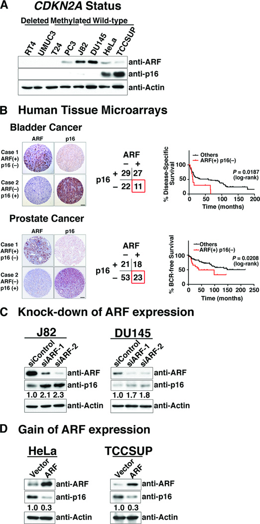
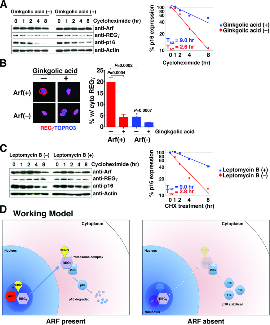
The cell-cycle regulatory gene INK4A-ARF (CDKN2A) has two alternative transcripts that produce entirely different proteins, namely p14(ARF) and p16, which have complementary functions as regulators of p53 and pRB tumor suppressor pathways, respectively. The unusual organization of INK4A-ARF has long led to speculation of a need for coordinated regulation of p14(ARF) and p16. We now show that p14(ARF) (ARF) regulates the stability of p16 protein in human cancer cell lines, as well as in mouse embryonic fibroblasts (MEFs). In particular, ARF promotes rapid degradation of p16 protein, which is mediated by the proteasome and, more specifically, by interaction of ARF with one of its subunits, REGγ. Furthermore, this ARF-dependent destabilization of p16 can be abrogated by knockdown of REGγ or by pharmacologic blockade of its nuclear export. Thus, our findings have uncovered a novel crosstalk of 2 key tumor suppressors mediated by a REGγ-dependent mechanism. The ability of ARF to control p16 stability may influence cell-cycle function.

Implications: The ability of ARF to control p16 stability may influence cell cycle function. Visual Overview: http://mcr.aacrjournals.org/content/current.


Conflict of interest statement

Disclosure: The authors disclose no potential conflicts of interest.

Figures

Figure 1
Figure 1
ARF regulates p16 protein levels in human cancer cells. (A) Inverse expression of ARF and p16 in human cancer cells. Western blot analyses of showing the expression levels of ARF and p16 proteins in the indicated human cancer cell lines, in which the CDNK2A gene is either deleted, methylated, or intact, as indicated. (B) Association of ARF and p16 expression with clinical outcome in bladder and prostate cancer. Representative images and categorical results of ARF and p16 immunostaining of tissue microarrays of human bladder and prostate cancer. Kaplan-Meier analyses show disease-specific survival of bladder cancer patients, and biochemical relapse (BCR)-free survival of prostate cancer patients. (C) Consequences of ARF knock-down for expression of p16 protein in J82 and DU145 cells using two independent ARF siRNA (or a scrambled siRNA as a control). (D) Consequences of expressing exogenous ARF in HeLa and TCCSUP cells following transfection with an ARF cDNA (or the empty vector as a control). In C and D the relative expression levels of p16 are indicated as determined using ImageJ software.
Figure 2
Figure 2
Arf regulates the stability of p16 protein via REGγ-dependent proteasome degradation in mouse embryonic fibroblasts. (A) Arf regulates p16 protein stability. Arf(+) and Arf(−) MEFs were treated with cycloheximide (50 µg/ml) for indicated time in hours. (Left) Western blot analyses showing relative protein expression levels. (Right) Relative change in p16 expression as a function of time showing the half-life (T1/2) was calculated from approximation curves. Note that in all approximation curves shown, the change in p16 expression is presented relative to the normalized expression levels (so it takes into account the change in basal levels in the cells). (B) Arf-mediated destablilization of p16 protein is counteracted by proteasome inhibitor. Arf(+) and Arf(−) MEFs were untreated or treated with cycloheximide (50 µg/ml) in the presence or absence of bortezomib (5 µM) and analyzed by Western blot analyses. (C) Arf interacts with REGγ in MEFs. (Left) Co-immunoprecipitation of endogenous Arf with endogenous REGγ using an anti-Arf antibody. (Right) Co-immunoprecipitation of exogenous HA-tagged REGγ with endogenous Arf using an anti-HA antibody. (D) REGγ is required for Arf-mediated destablization of p16 protein levels. Arf(+) MEFs were treated with treated two independent REGγ siRNA (or a scrambled siRNA as a control) followed by cycloheximide (50 µg/ml) for indicated time in hours. (Left) Western blot analyses showing relative protein expression levels. (Right) Relative change in p16 expression as a function of time showing the half-life (T1/2) was calculated from approximation curves. In A, B, and D the relative expression levels of p16 are indicated as determined using ImageJ software.
Figure 3
Figure 3
Arf-mediated destablization of p16 protein is mediated by nuclear export of REGγ. (A) Inhibition of SUMOylation stabilizes p16 expression in Arf-positive MEFs. Arf(+) MEFs were treated or untreated with ginkgolic acid (5 µM) for 4 hr followed by treatment with cycloheximide (50 µg/ml) for the indicated time in hours. (Left) Western blot analyses showing relative protein expression levels. (Right) Relative change in p16 expression as a function of time showing the half-life (T1/2) calculated from approximation curves. (B) Inhibition of SUMOylation reduces cytoplasmic localization of REGγ. Arf(+) and Arf(−) MEFs were transfected with an expression plasmid encoding HA-REGγ and treated with bortezomib (5 µM) and ginkgolic acid (5 µM) for 8 hr. (Left) Immunofluorescence images showing HA-REGγ localization in Arf(+) and Arf(−) MEFs detected using anti-HA antibody or detection of the nuclear marker TOPRO3. (Right) Percentage of cells in each condition having cytoplasmic expression of the REGγ. The chart summarize the results from 3 independent assays, each counting a minimum of 100 cells per variable. (C) Block of nuclear export of REGγ stabilizes p16 expression in Arf-positive MEFs. Arf(+) MEFs were treated or untreated with Leptomycin B (50 ng/ml) for 4 hr followed by treatment with cycloheximide (50 µg/ml) for the indicated time in hours. (Left) Western blot analyses showing relative protein expression levels. (Right) Relative change in p16 expression as a function of time showing the half-life (T1/2) calculated from approximation curves. (D) Working model. Discussed in the text. In A, and C the relative expression levels of p16 are indicated as determined using ImageJ software.

References

    1. Ruas M, Peters G. The p16INK4a/CDKN2A tumor suppressor and its relatives. Biochim Biophys Acta. 1998;1378:F115–F177. - PubMed
    1. Sharpless NE. INK4a/ARF: a multifunctional tumor suppressor locus. Mutat Res. 2005;576:22–38. - PubMed
    1. Kamijo T, Zindy F, Roussel MF, Quelle DE, Downing JR, Ashmun RA, et al. Tumor suppression at the mouse INK4a locus mediated by the alternative reading frame product p19ARF. Cell. 1997;91:649–659. - PubMed
    1. Quelle DE, Zindy F, Ashmun RA, Sherr CJ. Alternative reading frames of the INK4a tumor suppressor gene encode two unrelated proteins capable of inducing cell cycle arrest. Cell. 1995;83:993–1000. - PubMed
    1. Sherr CJ. The INK4a/ARF network in tumour suppression. Nature reviews Molecular cell biology. 2001;2:731–737. - PubMed

Publication types

MeSH terms

Substances