Review
. 2013 Jul;23(7):1069-80.
doi: 10.1101/gr.157586.113.

Insights into neural crest development and evolution from genomic analysis

Affiliations
Review

Insights into neural crest development and evolution from genomic analysis

Marcos Simões-Costa et al. Genome Res. 2013 Jul.

Abstract

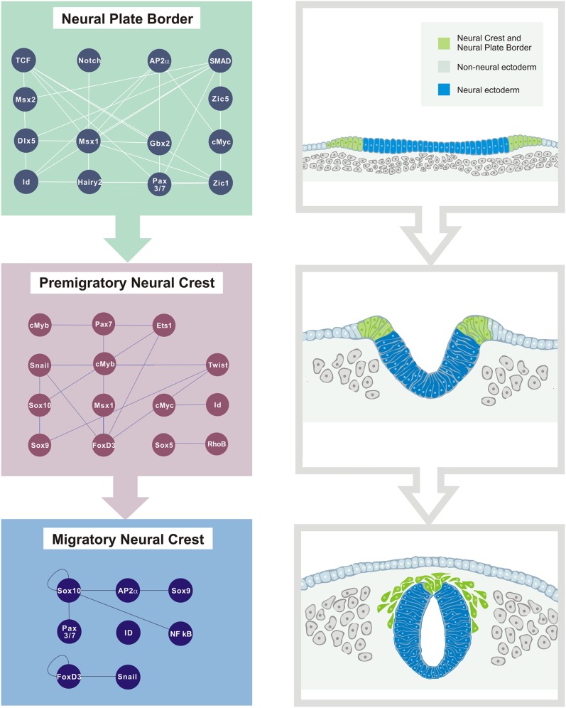
The neural crest is an excellent model system for the study of cell type diversification during embryonic development due to its multipotency, motility, and ability to form a broad array of derivatives ranging from neurons and glia, to cartilage, bone, and melanocytes. As a uniquely vertebrate cell population, it also offers important clues regarding vertebrate origins. In the past 30 yr, introduction of recombinant DNA technology has facilitated the dissection of the genetic program controlling neural crest development and has provided important insights into gene regulatory mechanisms underlying cell migration and differentiation. More recently, new genomic approaches have provided a platform and tools that are changing the depth and breadth of our understanding of neural crest development at a "systems" level. Such advances provide an insightful view of the regulatory landscape of neural crest cells and offer a new perspective on developmental as well as stem cell and cancer biology.


Figures

Figure 1.
Figure 1.
Morphogenetic movements during early neural crest development. (A) Schematic diagram of transverse sections through chick embryo during neurulation. Prospective neural crest cells reside in the neural plate border (green), a territory between the neural plate and the non-neural ectoderm. (B) As neurulation proceeds, the neural plate invaginates, resulting in the elevation of the neural folds, which contain neural crest precursors. (C) After neural tube closure, neural crest cells lose intercellular connections and undergo an epithelial to mesenchymal transition. Once they have delaminated from the neural tube, they migrate extensively to populate different niches throughout the embryo.
Figure 2.
Figure 2.
Contributions of different neural crest cell populations to adult tissues and organs. Depending on their axial level of origin and migratory pathway followed, neural crest cells adopt different fates and contribute to distinct tissues and organs. Cranial neural crest forms a large portion of the facial skeleton as well as cranial ganglia, most of the dental tissues, and the cornea. Vagal neural crest contributes to the valves and septa of the heart, the smooth muscle of the great vessels, and the enteric nervous system. Trunk neural crest gives rise to dorsal root and sympathetic ganglia of the peripheral nervous system and the chromaffin cells of the adrenal gland. Most caudally, the neural crest formed at the sacral region contributes to a small portion of the enteric nervous system. Melanocytes of the skin and integuments are derived from neural crest at all axial levels.
Figure 3.
Figure 3.
Cranial neural crest gene regulatory network (GRN). The neural crest gene GRN is composed of different regulatory modules arranged hierarchically. Each regulatory state defines cell identity and behavior at a given time and also drives the transition to the next level of the network. This is a simplified representation of the cranial neural crest GRN (Betancur et al. 2010a).
Figure 4.
Figure 4.
Strategies for cis-regulatory analysis of the neural crest. (A) Early approaches for identifying enhancers of neural crest genes included screening long stretches of noncoding DNA. Fragments from the locus of the gene of interest were cloned upstream of a minimal promoter and a reporter gene and tested in vivo by cell transfection or transgenesis. (B) Sequencing of vertebrate genomes facilitated the search for cis-regulatory modules. Computational comparison between different species reveals evolutionary conserved regions (ECRs) that are putative regulators of nearby genes. The ECRs are subsequently tested for activity by stable or transient transgenesis in different model organisms. (C) Novel approaches allow for genome-wide analysis of the cis-regulatory modules. Profiling of histone modification through ChIP-seq allows mapping of chromatin mark patterns and identification of active and poised enhancers. This method was used to annotate enhancers that are active in human neural crest cells induced from human embryonic stem cells (Rada-Iglesias et al. 2012). Once enhancers are validated in vivo, they become valuable tools that can be exploited in different contexts. (MP) Minimal promoter; (REP) reporter gene; (TF) transcription factor.

References

    1. Abitua PB, Wagner E, Navarrete IA, Levine M 2012. Identification of a rudimentary neural crest in a non-vertebrate chordate. Nature 492: 104–107 - PMC - PubMed
    1. Acloque H, Adams MS, Fishwick K, Bronner-Fraser M, Nieto MA 2009. Epithelial-mesenchymal transitions: The importance of changing cell state in development and disease. J Clin Invest 119: 1438–1449 - PMC - PubMed
    1. Adams MS, Gammill LS, Bronner-Fraser M 2008. Discovery of transcription factors and other candidate regulators of neural crest development. Dev Dyn 237: 1021–1033 - PMC - PubMed
    1. Agarwal P, Verzi MP, Nguyen T, Hu J, Ehlers ML, McCulley DJ, Xu SM, Dodou E, Anderson JP, Wei ML, et al. 2011. The MADS box transcription factor MEF2C regulates melanocyte development and is a direct transcriptional target and partner of SOX10. Development 138: 2555–2565 - PMC - PubMed
    1. Akiyama H, Chaboissier MC, Behringer RR, Rowitch DH, Schedl A, Epstein JA, de Crombrugghe B 2004. Essential role of Sox9 in the pathway that controls formation of cardiac valves and septa. Proc Natl Acad Sci 101: 6502–6507 - PMC - PubMed

Publication types