. 2013 Aug;16(8):1094-100.
doi: 10.1038/nn.3445. Epub 2013 Jun 30.

Cortical activation of accumbens hyperpolarization-active NMDARs mediates aversion-resistant alcohol intake

Affiliations

Cortical activation of accumbens hyperpolarization-active NMDARs mediates aversion-resistant alcohol intake

Taban Seif et al. Nat Neurosci. 2013 Aug.

Abstract

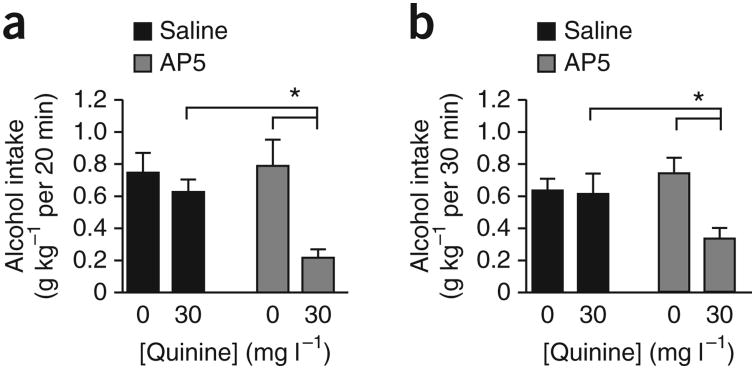
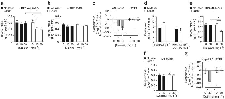
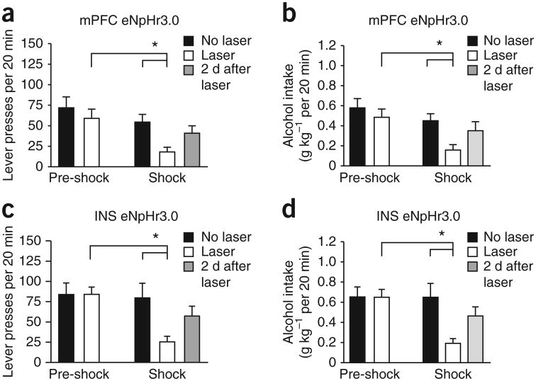
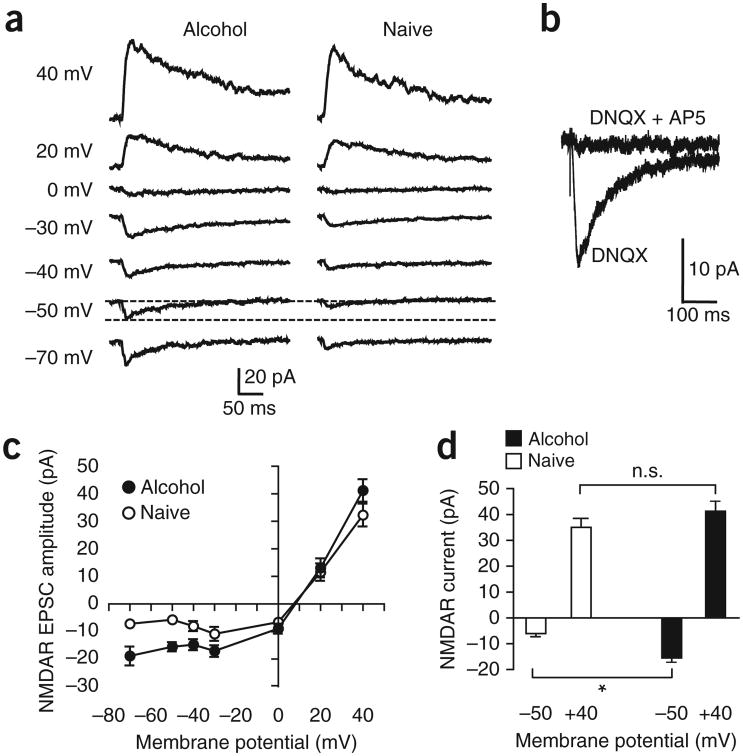
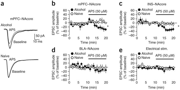
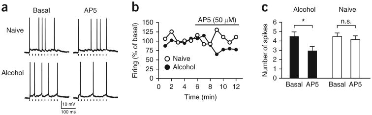
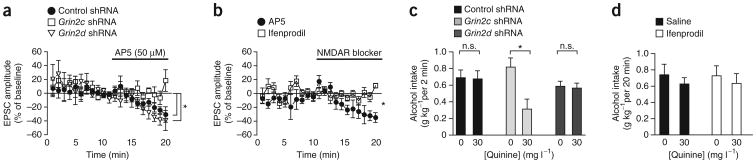
Compulsive drinking despite serious adverse medical, social and economic consequences is a characteristic of alcohol use disorders in humans. Although frontal cortical areas have been implicated in alcohol use disorders, little is known about the molecular mechanisms and pathways that sustain aversion-resistant intake. Here, we show that nucleus accumbens core (NAcore) NMDA-type glutamate receptors and medial prefrontal (mPFC) and insula glutamatergic inputs to the NAcore are necessary for aversion-resistant alcohol consumption in rats. Aversion-resistant intake was associated with a new type of NMDA receptor adaptation, in which hyperpolarization-active NMDA receptors were present at mPFC and insula but not amygdalar inputs in the NAcore. Accordingly, inhibition of Grin2c NMDA receptor subunits in the NAcore reduced aversion-resistant alcohol intake. None of these manipulations altered intake when alcohol was not paired with an aversive consequence. Our results identify a mechanism by which hyperpolarization-active NMDA receptors under mPFC- and insula-to-NAcore inputs sustain aversion-resistant alcohol intake.


Figures

Figure 1
Figure 1
Inhibition of NMDARs in the NAcore reduced aversion-resistant alcohol intake. (a) Intra-NAcore infusion of AP5 (0.5 μl per side of 1 μg μl−1) reduced intake, relative to that in saline-infused rats, of quinine-adulterated alcohol but not quinine-free alcohol in rats drinking alcohol 20 min d−1, 5 d week−1. (b) Similar results were observed when examining the impact of intra-NAcore AP5 on alcohol intake during the first 30 min of an intermittent overnight alcohol intake session (see Online Methods). Tukey post hoc *P < 0.05. Error bars indicate s.e.m.
Figure 2
Figure 2
Halorhodopsin inhibition of mPFC- and INS-to-NAcore inputs reduced quinine-resistant alcohol intake. (a) Halorhodopsin (eNpHr3.0) activation in mPFC-to-NAcore terminals significantly reduced intake, relative to that in the absence of laser stimulation, of alcohol adulterated with 10 or 30 mg l−1 quinine but not quinine-free alcohol. (b) Laser stimulation had no effect on alcohol intake in control rats infected with EYFP in mPFC-to-NAcore terminals. (c) To compare laser stimulation of halorhodopsin versus EYFP in mPFC terminals, we calculated the difference in intake between laser stimulation and no stimulation under six conditions: halorhodopsin + 10 or 30 mg l−1 quinine, halorhodopsin + no quinine, EYFP + 10 or 30 mg l−1 quinine, EYFP + no quinine. Laser stimulation only decreased alcohol intake with halorhodopsin + quinine. (d) Inhibiting mPFC-to-NAcore terminals did not alter intake of saccharin (sacc) ± quinine (quin). (e) Halorhodopsin inhibition of INS-to-NAcore terminals significantly reduced intake of quinine-adulterated alcohol (30 mg l−1 quinine) but not quinine-free alcohol. (f) Laser stimulation of INS-to-NAcore terminals had no effect in EYFP-infected rats. (g) Difference scores. Laser stimulation of INS-to-NAcore terminals decreased alcohol intake only with halorhodopsin + quinine. Tukey post hoc *P < 0.05. Error bars indicate s.e.m.
Figure 3
Figure 3
Halorhodopsin inhibition of mPFC- and INS-to-NAcore inputs reduced footshock-resistant alcohol intake. Rats underwent 7–8 weeks of fixed ratio 3 operant responding for alcohol, 20 min d−1, 5 d week−1. Thereafter, one in eight FR3 responses were paired with a footshock (0.25 mA, 0.5 s). By the sixth footshock session, alcohol intake could be considered shock-resistant in some rats (Supplementary Fig. 4a–c). We examined only those rats that gave more than 15 lever presses by the sixth footshock-paired session (10 of 17 rats for mPFC, 10 of 12 rats for INS). (a,b) Inhibiting mPFC-to-NAcore inputs significantly reduced responding for alcohol (a) and alcohol intake paired with intermittent footshock (b). Halorhodopsin (eNpHr3.0) had no effect on baseline, pre-shock sessions in this data set; however, when tested across all mPFC-injected rats, there was a small but significant reduction in baseline responding. (c,d) Inhibiting INS-to-NAcore inputs significantly reduced responding for alcohol (c) and alcohol intake paired with intermittent footshock (d). Halorhodopsin had no effect during baseline, pre-shock sessions. In addition, responding under footshock recovered in the 2 d after halorhodopsin exposure, suggesting no lasting impairments. “No laser” indicates average responding in the 2 d before halorhodopsin exposure under footshock. Tukey post hoc *P < 0.05. Error bars indicate s.e.m.
Figure 4
Figure 4
NAcore neurons from alcohol-drinking rats showed hyperpolarization-active NMDARs under mPFC-to-NAcore inputs. Currents were evoked by ChR2 stimulation of mPFC-to-NAcore terminals in alcohol-drinking and naive rats. (a) Example traces of NMDAR currents at different holding potentials. Dashed lines highlight the greater NMDAR current at −50 mV in a neuron from an alcohol-drinking rat. Not all voltages were determined for all neurons. (b) Example trace of AP5-sensitive NMDAR currents at −50 mV. (c) Grouped data for NMDAR currents under mPFC-to-NAcore inputs at different holding membrane potentials. For some data points the error bars fall within the symbol. The reversal potential for NMDARs was somewhat positive to zero mV without junction null correction, in line with adult mouse NAc shell. (d) Alcohol-drinking rats showed significantly greater NMDAR currents at −50 mV than alcohol-naive control rats, with no difference in NMDAR currents evoked at +40 mV holding potential. Tukey post hoc *P < 0.05; n.s., not significant. Error bars indicate s.e.m.
Figure 5
Figure 5
NAcore neurons from alcohol-drinking rats showed hyperpolarization-active NMDARs under mPFC and INS but not BLA inputs to the NAcore. (a,b) AP5 (50 μM) significantly reduced mPFC-ChR2-evoked EPSCs at a −70 mV membrane potential in alcohol-drinking but not control naive rats. (c) AP5-sensitive NMDAR currents were apparent in INS-ChR2-evoked EPSCs at −70 mV from alcohol-drinking but not control rats. (d,e) AP5 had no effect on EPSCs evoked at −70 mV by (d) ChR2 stimulation of BLA-NAcore terminals or (e) electrical field stimulation of glutamatergic terminals within the NAcore. *P < 0.05. Error bars indicate s.e.m.
Figure 6
Figure 6
NMDARs regulate evoked action potential firing under mPFC-to-NAcore inputs from alcohol-drinking but not naive rats. A ten-pulse train (20 Hz) of mPFC-ChR2-evoked EPSPs was stimulated every 10 s to evoke action potential firing; neurons were depolarized to about −55 mV (alcohol, −56.7 ± 2.5 mV; naive, −54.1 ± 2.9 mV; P = 0.491) with the patch amplifier to allow EPSPs to evoke ∼5 action potentials. (a,b) Traces (a) and time courses (b) from individual neurons showing AP5 inhibition of firing evoked by mPFC-to-NAcore ChR2 stimulation in neurons from alcohol-drinking but not naive rats. Each point represents the average of the six sweeps in that minute. Tick marks under firing traces in a indicate ChR2 stimulation. Firing does not completely track the timing of ChR2 stimulation, which likely reflects the strong potassium channel–mediated after hyperpolarization in NAcore neurons. (c) AP5 decreased firing in neurons from alcohol-drinking but not naive rats. Because of variability in firing across time, we averaged the firing across 4 min before AP5 administration (“basal”) and averaged the firing across the minutes 4–7 of AP5 exposure (“AP5”), as firing tended to run down in all cells after this point. For b, firing was normalized to the basal firing value. Tukey post hoc *P < 0.05. Error bars indicate s.e.m.
Figure 7
Figure 7
Grin2c but not Grin2d or Grin2b subunits mediate hyperpolarization-active NMDARs under mPFC-to-NAcore terminals and promote quinine-resistant alcohol intake. (a) Infection of NAcore neurons with Grin2c but not Grin2d or control shRNA prevented AP5 from inhibiting mPFC-evoked EPSCs at −70 mV. (b) The Grin2b inhibitor ifenprodil (2 μM) did not reduce mPFC-evoked EPSCs. Data for AP5 in b are the same as shown in Figure 5b. (c) Infecting NAcore neurons with Grin2c but not Grin2d or control shRNA reduced intake of quinine-adulterated alcohol. (d) Injecting the Grin2b inhibitor ifenprodil in the NAcore (1 μg μl−1) did not alter alcohol intake. *P < 0.05. Error bars indicate s.e.m.

Comment in

  • Drinking through the pain.
    Kash TL, Crabbe JC. Kash TL, et al. Nat Neurosci. 2013 Aug;16(8):987-8. doi: 10.1038/nn.3476. Nat Neurosci. 2013. PMID: 23887132 Free PMC article.

References

    1. Koob GF, Volkow ND. Neurocircuitry of addiction. Neuropsychopharmacology. 2010;35:217–238. - PMC - PubMed
    1. Naqvi NH, Bechara A. The insula and drug addiction: an interoceptive view of pleasure, urges, and decision-making. Brain Struct Funct. 2010;214:435–450. - PMC - PubMed
    1. Tiffany ST, Conklin CA. A cognitive processing model of alcohol craving and compulsive alcohol use. Addiction. 2000;95(suppl. 2):S145–S153. - PubMed
    1. Spanagel R. Alcoholism: a systems approach from molecular physiology to addictive behavior. Physiol Rev. 2009;89:649–705. - PubMed
    1. Wolffgramm J, Galli G, Thimm F, Heyne A. Animal models of addiction: models for therapeutic strategies? J Neural Transm. 2000;107:649–668. - PubMed

Publication types

MeSH terms