. 2013 Aug;16(8):1000-7.
doi: 10.1038/nn.3451. Epub 2013 Jun 30.

Arl13b-regulated cilia activities are essential for polarized radial glial scaffold formation

Affiliations

Arl13b-regulated cilia activities are essential for polarized radial glial scaffold formation

Holden Higginbotham et al. Nat Neurosci. 2013 Aug.

Abstract

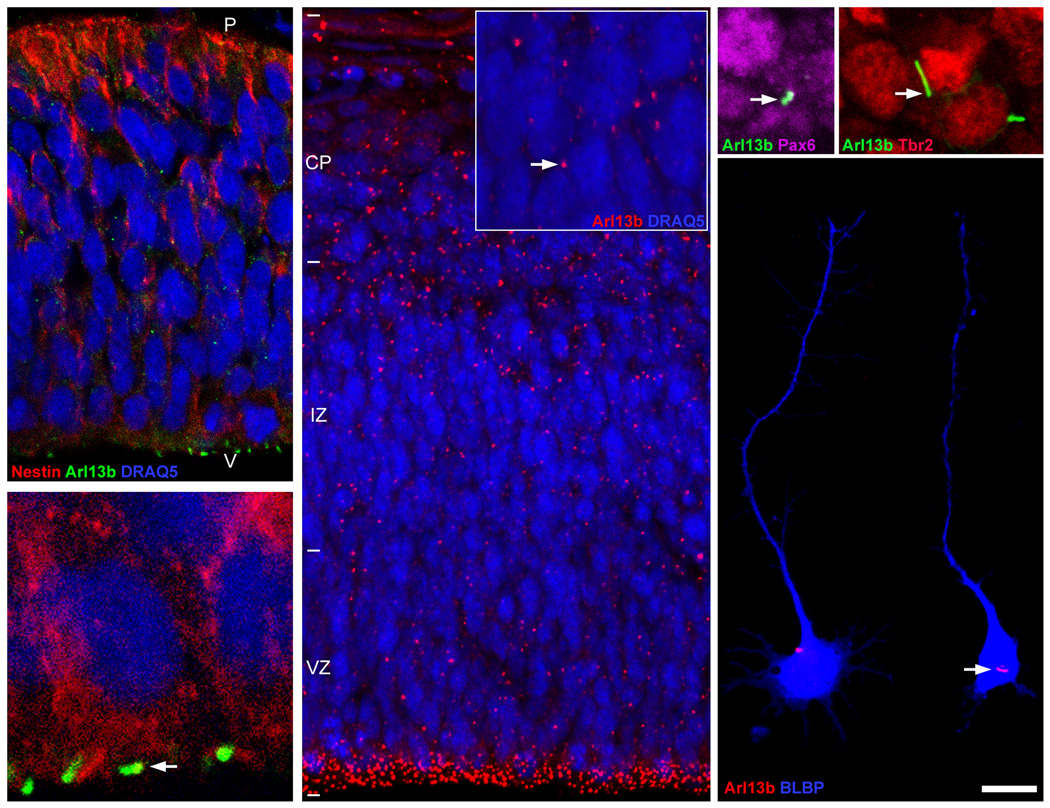
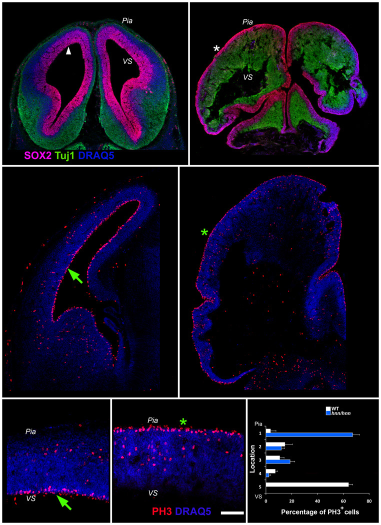
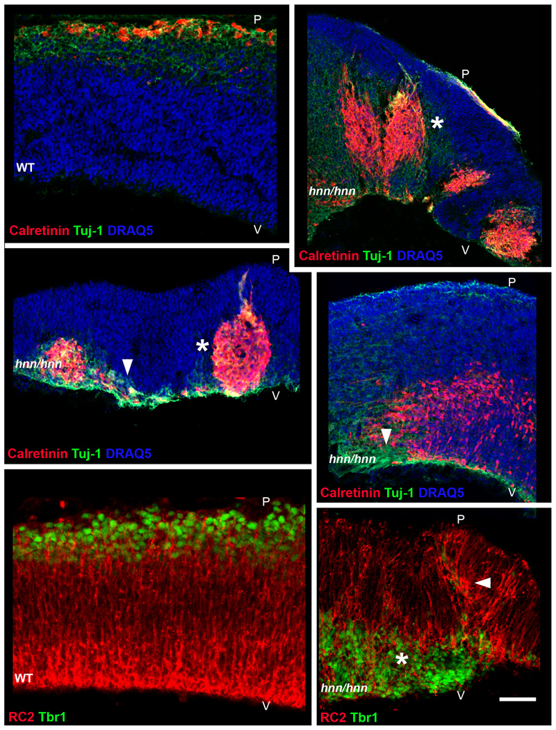
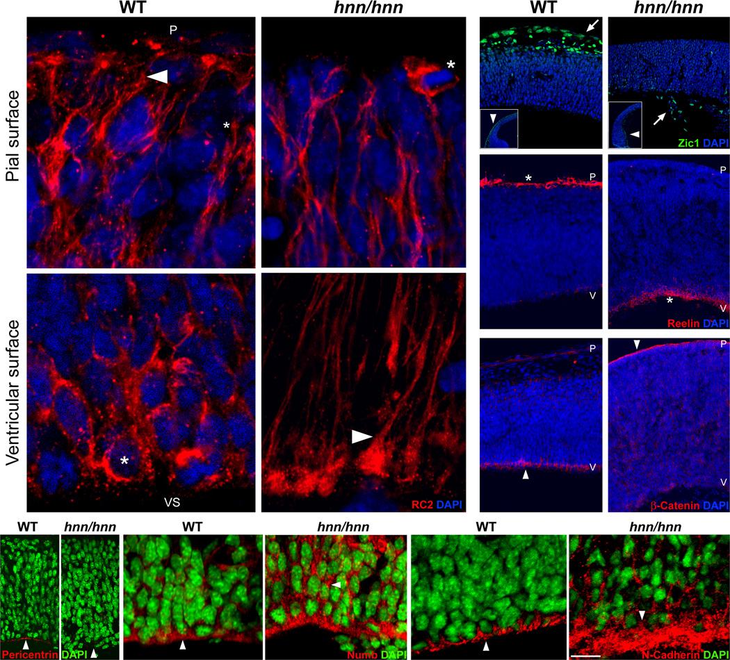
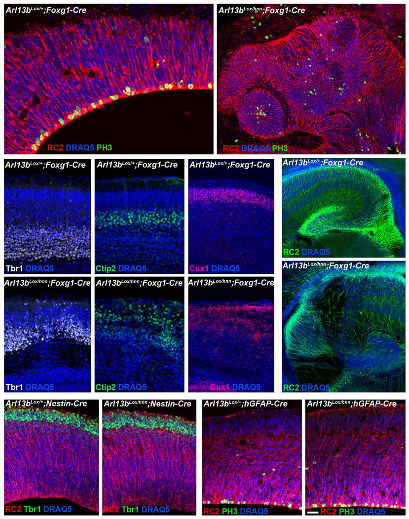
The construction of cerebral cortex begins with the formation of radial glia. Once formed, polarized radial glial cells divide either symmetrically or asymmetrically to balance appropriate production of progenitor cells and neurons. Following birth, neurons use the processes of radial glia as scaffolding for oriented migration. Radial glia therefore provide an instructive structural matrix to coordinate the generation and placement of distinct groups of cortical neurons in the developing cerebral cortex. We found that Arl13b, a cilia-enriched small GTPase that is mutated in Joubert syndrome, was critical for the initial formation of the polarized radial progenitor scaffold. Using developmental stage-specific deletion of Arl13b in mouse cortical progenitors, we found that early neuroepithelial deletion of ciliary Arl13b led to a reversal of the apical-basal polarity of radial progenitors and aberrant neuronal placement. Arl13b modulated ciliary signaling necessary for radial glial polarity. Our findings indicate that Arl13b signaling in primary cilia is crucial for the initial formation of a polarized radial glial scaffold and suggest that disruption of this process may contribute to aberrant neurodevelopment and brain abnormalities in Joubert syndrome-related ciliopathies.


Figures

Figure 1
Figure 1. Primary cilia in neuroepithelial cells and cortical progenitors
(A) Primary cilia (green dots) in a cross section of telencephalic neuroepithelium [E9] were labeled with anti-Arl13b antibodies. Neuroepithelial (red) cells were labeled with anti-nestin antibodies. (B) Higher magnification view of neuroepithelial cells with primary cilia (arrow). (C) Primary cilia (red dots) in a cross section of the developing cerebral wall [E14] were labeled with anti-Arl13b antibodies. Higher magnification inset (C) illustrates primary cilia (arrow) in VZ cells. (D–F) Different types of cortical progenitors were co-labeled with cell type-specific and anti-Arl13b antibodies. Radial progenitors (Pax6+[purple], D; BLBP+[blue], E) and intermediate progenitors (Tbr2+[red], F) possess primary cilia (arrow, D–F). Cells in A–C were nuclear counterstained (blue) with DRAQ5. Ventricular surface is towards the bottom in panels C (inset), D and F. Panels shown are representative examples from analysis of ten embryos per group. P, pial surface; V, ventricular surface; VZ, ventricular zone; IZ, intermediate zone; CP, cortical plate. Scale bar: A, 5 µm; B, D, F, 14 µm; C, 34 µm; E, 20 µm.
Figure 2
Figure 2. Disrupted formation of polarized radial progenitor scaffold in Arl13b mutants
(A) Loss of Arl13b and the resultant loss of primary cilia function lead to reversal of radial progenitor polarity. In E13 wild-type (WT) cortex, Sox2+ radial glial cell soma (purple, arrowhead) are localized near the ventricular surface (VS). In Arl13bhnn/hnn cortex, they are localized on the pial surface (asterisk, B). (C–F) Ectopic proliferation of radial progenitors in Arl13b mutants. Immunolabeling with anti-mitotic marker PH3 indicates that, compared to control (C, arrow), progenitors proliferate near the pial surface (D, asterisk) in Arl13bhnn/hnn cortex. Panels E and F are higher magnification views of the cerebral wall illustrating the proliferation of progenitors near the ventricular surface (arrow, E) and pial surface (asterisk, F) in WT and Arl13bhnn/hnn cerebral wall, respectively. (G) Normal interkinetic nuclear movement is required for the progenitors to migrate to the apical end (ventricular surface) to divide. Quantification of the location of proliferating progenitors in WT and Arl13bhnn/hnn cerebral wall indicates severe disruption of this process as most of the Arl13b mutant progenitors divide at the opposite end. Data shown are mean ± SEM (n=3 independent experiments; number of brains per group=3). Images shown are representative examples from analysis of 10 control and 10 mutant embryos. VS, ventricular surface; P, pial surface. Scale bar: A–B, 650 µm; C–D, 330 µm; E–F, 100 µm.
Figure 3
Figure 3. Disrupted cortical layer formation in Arl13b mutants
(A–D) The reversal of radial progenitor polarity affects neuronal migration and layer formation. Instead of the layer-like organization of different types of cortical neurons (red [Calretinin+]; green [Tuj-1+]) in E13 wild-type cortex (A), neurons in Arl13bhnn/hnn cortex (E13) migrate aberrantly and are placed ectopically (B–D). Neurons are frequently found near the ventricular surface (arrowheads, C, D) away from the cortical plate and often form tuber-like clusters (asterisk, B and C). (E–F) Co-immunolabeling of E13 WT (E) and mutant (F) cortices with radial progenitor-specific RC2 antibodies (red) and early generated, deeper layer neuronal marker Tbr1 (green) further illustrates the disrupted radial progenitor scaffold formation (arrowhead, F) and ectopic neuronal placement (asterisk, F) in Arl13b mutant cortex (F). Sections in A–D were nuclear counterstained (blue) with DRAQ5. Pial (P) and ventricular surface (V) are indicated. Images shown are representative examples from analysis of 10 control and 10 mutant embryos. Scale bar: A–D, 275 µm; E–F, 130 µm.
Figure 4
Figure 4. Cortical developmental disruptions in Arl13b mutants
(A–D) Aberrant localization of RC2+ radial glial endfeet near the ventricular surface. Consistent with the reversed polarity of radial progenitors, the normal localization of radial progenitor endfeet near the pial surface (arrowhead, A) and cell soma near the ventricular surface (asterisk, C) are reversed in E13 Arl13b mutants (B, D). (E–F) Disrupted formation of Zic1+ pial membrane in mutants. Compared to the WT pia (E, arrow), mutant (F, arrow) pia is discontinuous and misplaced. Inset panel (E) indicates normal cortical covering by pial membrane (arrowhead), whereas mutant pia (arrowhead, F [inset]) is discontinuous and often found on the opposite side of the cerebral wall. (G–P) Altered apical-basal organization of the cerebral cortex in mutants. (G–H) Reelin+ Cajal-Retzius neurons are normally found at the top of the cortex in layer 1 (asterisk, G). In contrast, they were localized to the apical surface (asterisk, H), near the ventricles in mutants. (I–J) Anti-β-catenin prominently labels the apical surface of progenitors (arrowhead, I), adjacent to the ventricles in WT cortex. In contrast, β-catenin labeling is evident in the opposite, basal surface (arrowhead, J) in mutants. Further, apical localizations of pericentrin (K), Numb (M) and N-Cadherin (O) are disrupted in mutants (L, N, P; compare arrowheads). P, pial surface; V, ventricular surface. Images are representative examples from analysis of 7 control and 7 mutant embryos. Scale bar: A–D, 17 µm; E–F, 110 µm; G, I, 190 µm; H, J, 300 µm; K–L, 60 µm; M–P, 30 µm.
Figure 5
Figure 5. Arl13b deletion in neuroepithelial cells disrupts radial progenitor scaffold organization and laminar organization of neurons in cerebral cortex
(A–B) Normal radial progenitor scaffold (RC2+) and progenitor proliferation (PH3+) in E13 control cerebral wall. (B) Loss of polarized radial progenitor organization and ectopic proliferation in E13 Arl13blox/hnn; Foxg1-Cre cortex following deletion of Arl13b in neuroepithelial cells. (C–H) Deeper and upper-layer neurons in E16 cortices from control and Arl13blox/hnn; Foxg1-Cre mice were labeled with anti-Tbr1, -Ctip2 (Bcl11b) and -Cux1 antibodies. Characteristic laminar organization of different classes of neurons was evident in control cortices (C–E), but was disrupted in Arl13blox/hnn; Foxg1-Cre cortex (F–H). (I–J) Hippocampus, which also contains polarized radial progenitors (I, green), does not form properly in Arl13blox/hnn; Foxg1-Cre mice (J). (K–N) Deletion of Arl13b following the formation of polarized radial progenitor scaffold does not affect radial progenitors. Nestin-Cre and hGFAP-Cre lines were used to ablate Arl13b from E10.5 and E13.5 radial progenitors, respectively. (K–L) In Arl13blox/hnn; Nestin-Cre cortex (L), RC2+ radial progenitors form normally and neuronal migration and placement resembles control cortex (K). (M–N) Similarly, radial progenitor scaffold is not disrupted in Arl13blox/hnn; hGFAP-Cre cortex (N), and progenitor proliferation is identical to that of control cortex (M). Early-born neurons (K–L) and mitotic progenitors (M–N) were labeled with anti-Tbr1 and anti-PH3 antibodies, respectively. Nuclei were counterstained with DAPI. Images are representative examples from analysis of 10 control and 10 mutant mice per group. Scale bar: A, 40 µm; B, 90 µm; C–H, 75 µm; I–J, 300 µm; K–N, 725 µm.
Figure 6
Figure 6. Altered primary cilia dynamics in Arl13b mutants
(A, B). Time-lapse imaging of 5-Htr6-labeled cilia illustrates that in WT progenitors (A), primary cilium changes its length, shape, orientation, and sometimes forms branches (arrowhead, A). In contrast, such dynamism is absent in Arl13b mutant cilium (B). Time interval between each panel is 6 minutes. Scale bar: 1 µm.
Figure 7
Figure 7. Disrupted localization and signalling of apical complex receptors Igf1R inArl13bhnn/hnn cortex
Immunolabeling of WT and mutant cortices with anti-Igf1R antibodies indicate disrupted localization of Igf1R in apical domains of the radial progenitors following Arl13b deletion (compare arrows in panels A and B). Insets (A, B) show ciliary localization of Igf1R. Ciliary Igf1R signalling leads to phosphorylation of Akt (C, arrow) and accumulation of phosphor-IRS (G, arrow) at the base of the cortical progenitor cilium. Arl13b deletion disrupts these characteristic signs of Igf1R signalling (D, F, H, J). Re-expression of Arl13b in Arl13b mutant primary cilia rescues these deficits (E [arrow], F, I [arrow], J). P-Akt, P-IRS-1 index indicates percentage of cilia with p-Akt or P-IRS-1 accumulation at the base of the cilium. Data shown are mean ± SEM. Number of cilia analyzed: p-AKT group-WT (77), hnn/hnn (93), hnn/hnn+Arl13b (69); p-IRS-1 group- WT (93), hnn/hnn (96), hnn/hnn+Arl13b (88). * and **, significant when compared with WT or hnn/hnn, respectively, at p < 0.05 (One way ANOVA with Tukey-Kramer test). Cells were obtained from four different litters. Cilia (A, B, C, D, G, H) were labeled with anti-ACIII antibodies. Scale bar: A–B, 18 µm; C–I, 1 µm.

References

    1. Rakic P. Specification of cerebral cortical areas. Science. 1988;241:170–176. - PubMed
    1. Miyata T, Kawaguchi A, Okano H, Ogawa M. Asymmetric inheritance of radial glial fibers by cortical neurons. Neuron. 2001;31:727–741. - PubMed
    1. Noctor SC, Martinez-Cerdeno V, Ivic L, Kriegstein AR. Cortical neurons arise in symmetric and asymmetric division zones and migrate through specific phases. Nat Neurosci. 2004;7:136–144. - PubMed
    1. Gotz M, Huttner WB. The cell biology of neurogenesis. Nat Rev Mol Cell Biol. 2005;6:777–788. - PubMed
    1. Manzini MC, Walsh CA. What disorders of cortical development tell us about the cortex: one plus one does not always make two. Curr Opin Genet Dev. 2011;21:333–339. - PMC - PubMed

Publication types

MeSH terms

Supplementary concepts