. 2013 Oct;94(4):835-43.
doi: 10.1189/jlb.1112566. Epub 2013 Jul 1.

Cocaine exposure enhances permissiveness of quiescent T cells to HIV infection

Affiliations

Cocaine exposure enhances permissiveness of quiescent T cells to HIV infection

Sohn G Kim et al. J Leukoc Biol. 2013 Oct.

Abstract

In vivo and in vitro exposure to stimulants has been associated with increased levels of HIV infection in PBMCs. Among these lymphocyte subsets, quiescent CD4(+) T cells make up the majority of circulating T cells in the blood. Others and we have demonstrated that HIV infects this population of cells inefficiently. However, minor changes in their cell state can render them permissive to infection, significantly impacting the viral reservoir. We have hypothesized that stimulants, such as cocaine, may perturb the activation state of quiescent cells enhancing permissiveness to infection. Quiescent T cells isolated from healthy human donors were exposed to cocaine and infected with HIV. Samples were harvested at different time-points to assess the impact of cocaine on their susceptibility to infection at various stages of the HIV life cycle. Our data show that a 3-day exposure to cocaine enhanced infection of quiescent cells, an effect that appears to be mediated by σ1R and D4R. Overall, our results indicate that cocaine-mediated effects on quiescent T cells may increase the pool of infection-susceptible T cells. The latter underscores the impact that stimulants have on HIV-seropositive individuals and the challenges posed for treatment.

Keywords: human immunodeficiency virus; life cycle; reservoir; stimulant abuse; σ1 and D4 receptors.


Figures

Figure 1.
Figure 1.. Cocaine treatment of quiescent T cells induces phenotypic changes.
Quiescent T cells (Quiescent) were exposed to cocaine (Cocaine) for 3 days or stimulated with anti-CD3/anti-CD28 (CD3/CD28). Cells were then harvested and analyzed by flow cytometry for cell cycle progression and surface marker expression changes. (A) For cell cycle progression, cells were stained with 7-AAD (DNA) and Pyronin Y (RNA), as shown in the upper panels from one representative donor. The increased entry into G1b, following a 3-day cocaine treatment, is statistically significant, as shown in the lower bar graph (n=7; **P<0.01, one-tailed Student's t-test). Cocaine treatment had no negative effect on cell viability. (B) Cells were also assessed for the expression of CCR5 (n=8; **P<0.01, one-tailed Student's t-test) and CXCR4 (not significant), as well as (C) T cell activation markers (not significant between Quiescent and Cocaine groups).
Figure 2.
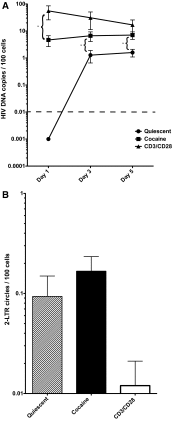
Figure 2.. Cocaine treatment of quiescent T cells results in increased levels and kinetics of reverse transcription.
Quiescent cells were exposed to cocaine for 3 days. Cells were then infected and harvested at different time-points postinfection. Samples were used to assess the kinetics of HIV infection in cocaine-treated cells compared with quiescent as well as prestimulated T cells. (A) Cells (0.3×10⁶) were harvested at different time-points after infection, and DNA was extracted for viral DNA analysis. DNA extraction and real-time PCR analysis were performed. The limit of detection (indicated by a dashed line) was 0.01% cells, based on AZT-treated negative controls (for Day 1, *P<0.05 Cocaine vs. CD3/CD28; for Days 3 and 5, *P<0.05 Quiescent vs. Cocaine, Student's t-test). (B) Quantitation of 2-LTR circle formation in cocaine treated, untreated and activated T cells on day after infection using real time PCR. The data shown are the average from three representative donors (out of 5).
Figure 3.
Figure 3.. Integrated HIV levels are higher in cocaine-treated quiescent T cells.
Integration of HIV DNA was assessed by an Alu-based integration assay. The limit of detection was 0.01% cells. In the control experiments, there was no background from nonintegrated viral DNA, and values for the integrated viral DNA varied <20% within triplicates (for Day 1, *P<0.05 Cocaine vs. CD3/CD28; for Days 3 and 5, *P<0.05 Quiescent vs. Cocaine, Student's t-test). The data shown are the average from three representative donors (out of five).
Figure 4.
Figure 4.. Viral mRNA and protein expression increased in cocaine-treated quiescent cells.
(A) Total cellular RNA was isolated from all groups and used to detect the amount of multiply spliced (tat/rev) viral RNA. (B) All cell groups were infected with HIV at a MOI of 1 following treatment. Cells were then harvested at various times and stained with PE-conjugated anti-Kc57 (RD1; log10 fluorescence), a Gag p24-specific antibody. The panel of flow cytometry dot plots is from one representative donor, 3 days postinfection. The data shown are the average from three representative donors (out of five; *P<0.05).
Figure 5.
Figure 5.. Cocaine effects are mediated via the DRD4R.
(A) Quiescent CD4 T cells from four donors (**P<0.01; ***P<0.001) were stained for antibodies against the σ1R and DRD4R (see Materials and Methods) and analyzed by flow cytometry, as shown on the scatter dot plots. The groups labeled Staining control are cells incubated with secondary antibody alone. Histogram panels are from one representative donor. The dashed-line histograms are cells incubated with secondary antibody alone. (B) Quiescent CD4 T cells were treated with cocaine, D4R, or σ1R agonists for 3 days. A subset of the cocaine-treatment group was pretreated with DRD4R or σ1R antagonists, 4 h prior to cocaine exposure. The reagents used are: CP-226,269 (DRD4R agonist at 32 nM), L-687,384 (σ1R agonist at 40 μM), L-745,870 (DRD4R antagonist at 0.1 μΜ), and BD1047 (σ1R antagonist at 1 μΜ). Cells treated with cocaine or the D4 agonist resulted in increased CCR5 expression. The data are the average from three different donors (left, *P<0.05; **P<0.01). Treated cells were also infected with HIV-189.6 and assayed for p24(Gag) expression. D4 agonist-treated cells mimicked the cocaine-induced permissiveness to infection. The data indicate the fold increase of percent p24-positive cells over cocaine only-treated cells and are the average from four different donors (right, *P<0.05 D4 antagonist vs. Cocaine and D4 Agonist vs. Cocaine).

References

    1. Zack J. A., Arrigo S. J., Weitsman S. R., Go A. S., Haislip A., Chen I. S. (1990) HIV-1 entry into quiescent primary lymphocytes: molecular analysis reveals a labile, latent viral structure. Cell 61, 213–222 - PubMed
    1. Vatakis D. N., Bristol G., Wilkinson T. A., Chow S. A., Zack J. A. (2007) Immediate activation fails to rescue efficient human immunodeficiency virus replication in quiescent CD4+ T cells. J. Virol. 81, 3574–3582 - PMC - PubMed
    1. Swiggard W. J., O'Doherty U., McGain D., Jeyakumar D., Malim M. H. (2004) Long HIV type 1 reverse transcripts can accumulate stably within resting CD4+ T cells while short ones are degraded. AIDS Res. Hum. Retroviruses 20, 285–295 - PubMed
    1. Zhou Y., Zhang H., Siliciano J. D., Siliciano R. F. (2005) Kinetics of human immunodeficiency virus type 1 decay following entry into resting CD4+ T cells. J. Virol. 79, 2199–2210 - PMC - PubMed
    1. Cole S. W., Jamieson B. D., Zack J. A. (1999) cAMP up-regulates cell surface expression of lymphocyte CXCR4: implications for chemotaxis and HIV-1 infection. J. Immunol. 162, 1392–1400 - PubMed

Publication types