. 2013 Sep;81(9):3414-25.
doi: 10.1128/IAI.00784-12. Epub 2013 Jul 1.

Donor CD8+ T cells prevent Toxoplasma gondii de-encystation but fail to rescue the exhausted endogenous CD8+ T cell population

Affiliations

Donor CD8+ T cells prevent Toxoplasma gondii de-encystation but fail to rescue the exhausted endogenous CD8+ T cell population

Rajarshi Bhadra et al. Infect Immun. 2013 Sep.

Abstract

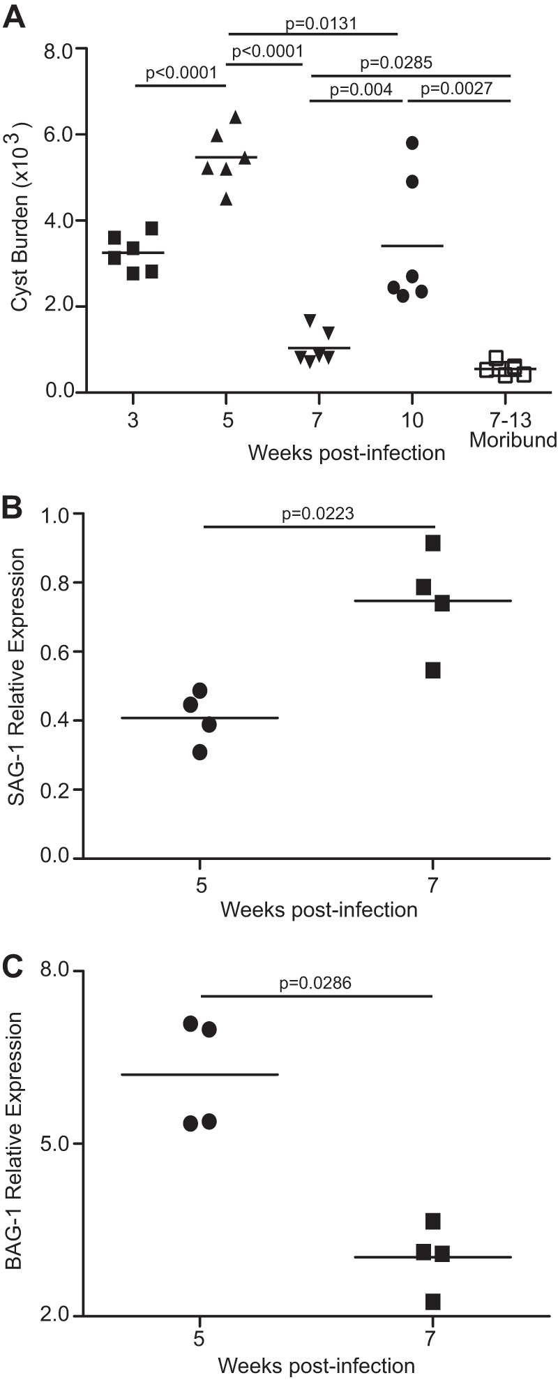
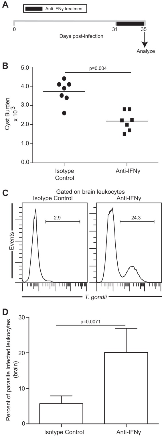
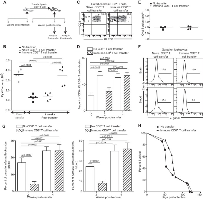
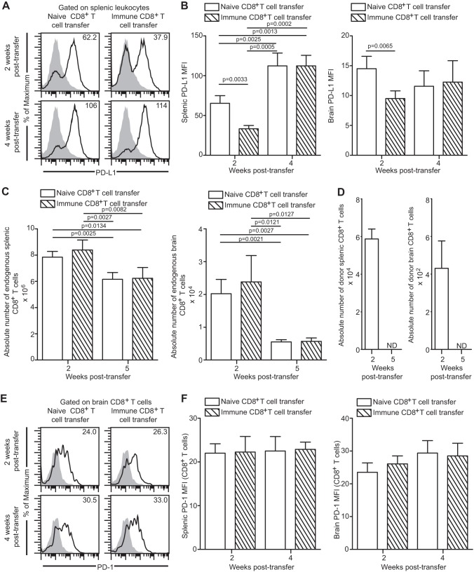
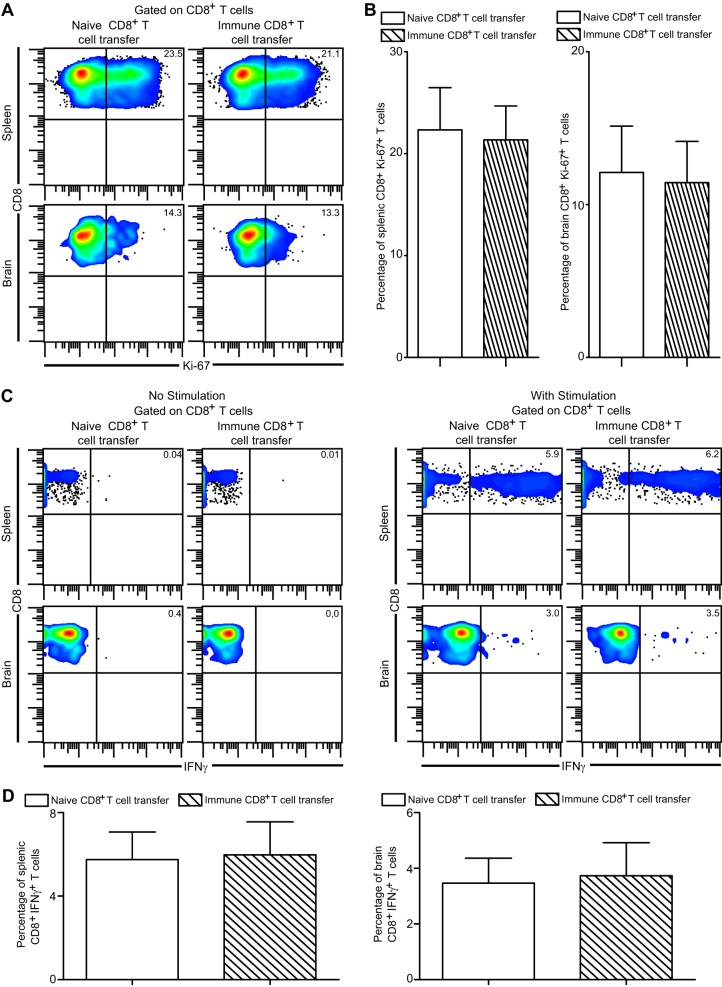
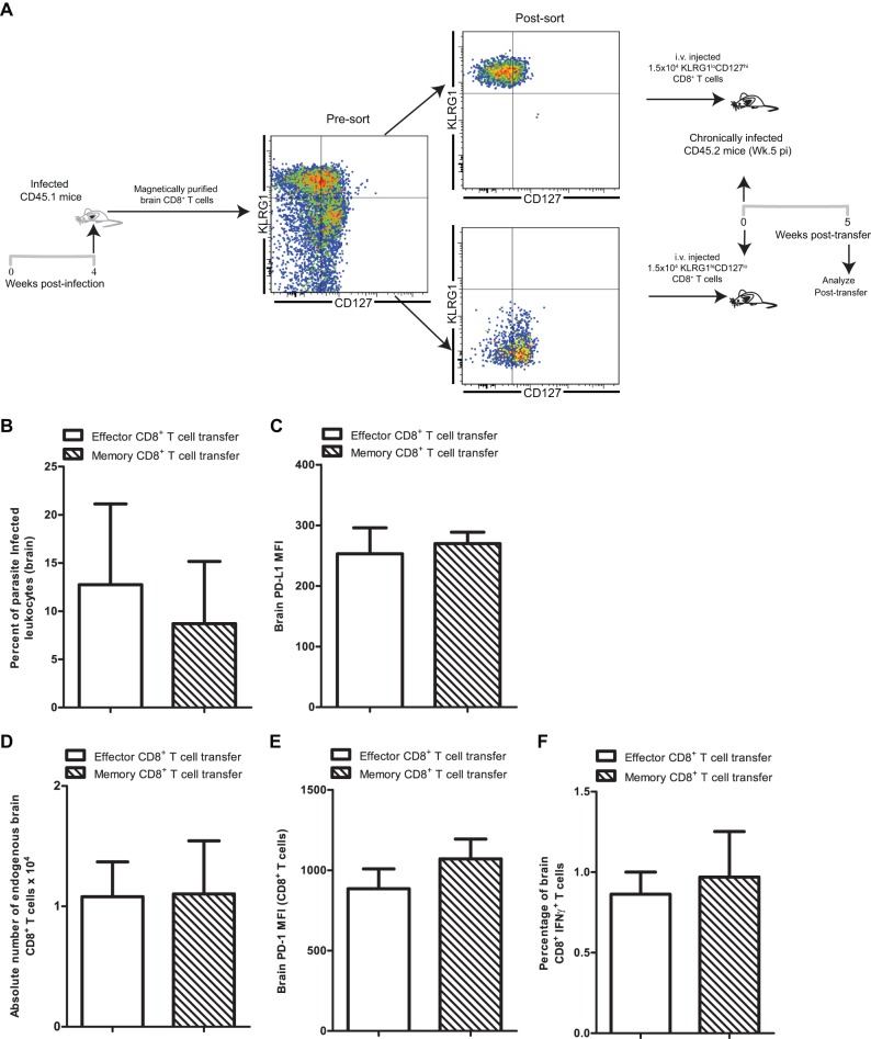
Functional exhaustion of CD8(+) T cells due to increased expression of inhibitory molecule PD-1 (Programmed Death-1) causes reactivation of latent disease during later phases of chronic toxoplasmosis. Onset of disease recrudescence results in decreased parasite cyst burden concomitant with parasites undergoing stage conversion from a primarily encysted, quiescent bradyzoite to a fast-replicating, highly motile tachyzoite. Thus, reduced cyst burden is one of the early hallmarks of disease recrudescence. This was further validated by depleting gamma interferon (IFN-γ), a cytokine known to control latent toxoplasmosis, in chronically infected prerecrudescent mice. Since CD8(+) T cells (an important source of IFN-γ) lose their functionality during the later phases of chronic toxoplasmosis, we next examined if adoptive transfer of functional CD8(+) T cells from acutely infected donors to the chronically infected prerecrudescent hosts could impede parasite de-encystation and rescue exhausted CD8(+) T cells. While the transfer of immune CD8(+) T cells temporarily restricted the breakdown of cysts, the exhausted endogenous CD8(+) T cell population was not rescued. Over time, the donor population got deleted, resulting in parasite de-encystation and host mortality. Considering that donor CD8(+) T cells fail to become long-lived, one of the cardinal features of memory CD8(+) T cells, it bears the implication that memory CD8 differentiation is impaired during chronic toxoplasmosis. Moreover, our data strongly suggest that while adoptive immunotherapy can prevent parasite de-encystation transiently, reduced antigen burden in the chronic phase by itself is insufficient for rescue of exhausted CD8(+) T cells. The conclusions of this study have profound ramifications in designing immunotherapeutics against chronic toxoplasmosis.


Figures

Fig 1
Fig 1
Decreased cyst burden during late-chronic T. gondii infection correlates with parasite reactivation. (A) C57BL/6 mice were infected with 10 ME49 cysts orally, and the number of brain cysts was enumerated at weeks 3, 5, 7, and 10 postinfection. A similar trend in cyst kinetics was observed in 3 independent experiments. (B and C) SAG-1 and BAG-1 relative expression levels were measured in brain at week 7 pi. The transcript level at day 10 was taken as 1. The data represent at least two experiments with at least 4 mice per group. Error bars represent standard deviations throughout.
Fig 2
Fig 2
Reduced cyst burden and increased leukocyte parasitization in mice treated with αIFN-γ during early-chronic infection. (A) αIFN-γ antibody was injected daily, starting at day 31 pi (n = 7). Animals were sacrificed 4 days after initiation of treatment. (B) The number of brain cysts was enumerated in isotype- or αIFN-γ antibody-treated mice. (C and D) T. gondii-infected cells in brain leukocyte-gated samples (n = 5) were assayed by flow cytometry, and the results are depicted as histograms (C) or a bar graph (D). The data represent two experiments with at least 4 mice per group.
Fig 3
Fig 3
Control of parasite reactivation by adoptive transfer of immune CD8+ T cells. (A) Splenic CD8+ T cells (4 × 10⁶) purified from donor mice at week 2 pi were adoptively transferred via the i.v. route into chronically infected recipient mice at week 5 pi. (B) Cyst burden in brains of recipient mice (n = 5) was evaluated 2 weeks posttransfer. (C and D) Frequencies of KLRG1-expressing recipient CD8+ T cells (n = 4) in brain are shown as density plots (C) (2 weeks posttransfer) or as a bar graph (D). (E) Cyst burden in brains of recipient mice was assessed at 4 weeks posttransfer. (F and G) T. gondii-infected cells in brain and blood leukocyte-gated samples from recipient mice (n = 5) were assayed by flow cytometry. (F) A histogram depicts frequency of parasite-infected leukocytes at 2 weeks posttransfer. (G) A bar graph (left panel, brain; right panel, blood) shows percentages of infected leukocytes at weeks 2 and 4 posttransfer in recipient animals that received immune CD8+ T cell transfer or PBS. (H) Survival of immune CD8+ T cell transfer recipients (n = 8) and saline-treated controls (n = 7) was monitored on a daily basis. Data represent two experiments.
Fig 4
Fig 4
Donor CD8+ T cells do not ameliorate endogenous CD8+ T cell exhaustion during late-chronic toxoplasmosis. (A and B) PD-L1 expression was assayed on total splenic and brain leukocytes by flow cytometry at weeks 2 and 4 posttransfer. Data are presented as representative histograms (A) or bar graphs (B). (C) The absolute number of endogenous CD8+ T cells in spleen and brain in the recipient mice was computed. (D) A bar graph represents the absolute number of donor CD8+ T cells in spleen (left panel) and brain (right panel) in immune CD8+ T cell transfer recipients. N.D. denotes “not detectable.” (E and F) PD-1 expression on endogenous CD8+ T cells in spleen and brain was evaluated by flow cytometry. Data are presented as a histogram (E) or as a bar graph (F) (left panel, spleen; right panel, brain). The data represent 2 experiments with at least 4 mice per group.
Fig 5
Fig 5
Donor CD8+ T cells fail to rescue endogenous CD8+ T cell functionality during late-chronic toxoplasmosis. (A and B) In vivo endogenous CD8+ T cell proliferation was assessed in spleen and brain of recipient animals at 2 weeks posttransfer by measuring Ki-67 expression via intracellular staining. Data are presented as pseudocolor plots (A) or as bar graphs (B) (left panel, spleen; right panel, brain). (C and D) Splenocytes and brain mononuclear cells were stimulated with TLA as described in Materials and Methods and then evaluated for IFN-γ production by endogenous CD8+ T cells at 2 weeks posttransfer. Data are depicted as a pseudocolor plot (C) (left panel, unstimulated; right panel, TLA stimulated) or as bar graphs (D) (left panel, spleen; right panel, brain). The data are representative of 2 experiments with 3 to 4 mice per group.
Fig 6
Fig 6
Donor memory CD8+ T cells fail to rescue endogenous CD8+ T cells during late-chronic toxoplasmosis. (A) Donor CD45.1 mice were infected with T. gondii. Four weeks later, brain CD8+ T cells were purified from donor CD45.1 mice and subsequently sorted into effector and memory T cell subsets. Sorted cells (1.5 × 10⁴) were transferred into chronically infected CD45.2 recipient mice (n = 4) at week 5 postinfection via the i.v. route. (B) T. gondii-infected cells in brain leukocyte-gated samples from recipient mice were assayed by flow cytometry. A bar graph shows percentages of parasite-infected leukocytes at 5 weeks posttransfer (10 weeks postinfection) in recipient animals that received effector CD8+ T cells or memory CD8+ T cells. (C) PD-L1 expression was assayed on total brain leukocytes by flow cytometry at week 5 posttransfer in recipient animals. (D) A bar graph represents the absolute number of endogenous CD8+ T cells in brain of CD8+ T cell recipients. (E) PD-1 expression on endogenous CD8+ T cells in brain was evaluated by flow cytometry. (F) Brain mononuclear cells were stimulated with TLA and then evaluated for IFN-γ production by endogenous CD8+ T cells at 5 weeks posttransfer. Data were acquired using a Cytek-upgraded 8-color BD FACSCalibur flow cytometer which accounts for differences in fluorescence scale.

References

    1. Kur J, Holec-Gasior L, Hiszczynska-Sawicka E. 2009. Current status of toxoplasmosis vaccine development. Expert Rev. Vaccines 8:791–808 - PubMed
    1. Falusi O, French AL, Seaberg EC, Tien PC, Watts DH, Minkoff H, Piessens E, Kovacs A, Anastos K, Cohen MH. 2002. Prevalence and predictors of Toxoplasma seropositivity in women with and at risk for human immunodeficiency virus infection. Clin. Infect. Dis. 35:1414–1417 - PMC - PubMed
    1. Grant IH, Gold JW, Rosenblum M, Niedzwiecki D, Armstrong D. 1990. Toxoplasma gondii serology in HIV-infected patients: the development of central nervous system toxoplasmosis in AIDS. AIDS 4:519–521 - PubMed
    1. Bhadra R, Gigley JP, Khan IA. 2011. The CD8 T-cell road to immunotherapy of toxoplasmosis. Immunotherapy 3:789–801 - PMC - PubMed
    1. Gigley JP, Bhadra R, Khan IA. 2011. CD8 T cells and Toxoplasma gondii: a new paradigm. J. Parasitol. Res. 2011:243796.10.1155/2011/243796 - DOI - PMC - PubMed

Publication types

Substances