

Review
doi: 10.1152/physiol.00006.2013.
Elucidating immune mechanisms causing hypertension during pregnancy
Affiliations
- PMID: 23817797
- PMCID: PMC3742131
- DOI: 10.1152/physiol.00006.2013
Item in Clipboard
Review
Elucidating immune mechanisms causing hypertension during pregnancy
Physiology (Bethesda).
2013 Jul.
Abstract
Preeclampsia is associated with hypertension and increased infant and maternal morbidity and mortality. The underlying cause of preeclampsia is largely unknown, but it is clear that an immunological component plays a key pathophysiological role. This review will highlight immunological key players in the pathology of preeclampsia and discuss their role in the pathophysiology observed in the reduced placental perfusion (RUPP) rat model of preeclampsia.
Conflict of interest statement
No conflicts of interest, financial or otherwise, are declared by the author(s).
Figures
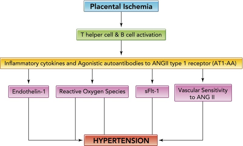
Hypertension in response to placental ischemia Hypertension in response to placental ischemia proceeds via immune activation, CD4+ T-cells mediating the release of angiotensin II type-1 receptor autoantibody (AT1-AA), and inflammatory cytokines that contribute to the increased vasoactive peptide ET-1 increased sensitivity to ANGII, oxidative stress, and sFlt-1, all known players in the pathophysiology of preeclampsia.
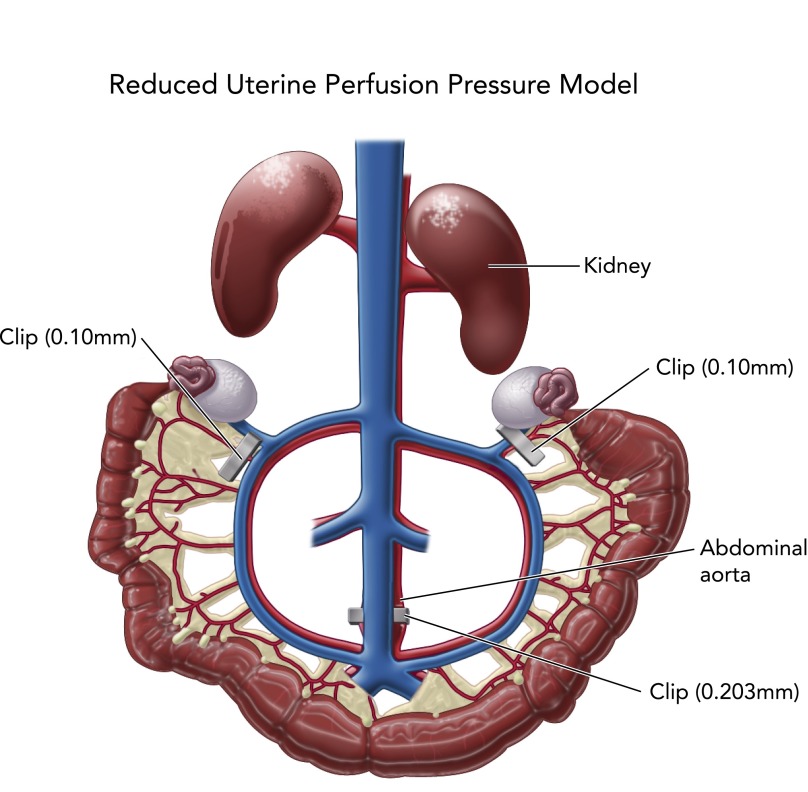
Reduced uterine perfusion pressure model Reduced uterine perfusion pressure model is utilized to induce placental ischemia in pregnant rats on day 14 of gestation; blood pressure and soluble factors are collected on day 19 of gestation.
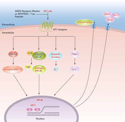
Signal cascades of the angiotensin II type 1 receptor autoantibodies The agonistic angiotensin II type 1 receptor autoantibodies (AT1-AA) induce signaling by the angiotensin II type 1 receptor (AT1-receptor), inhibited by AT1-receptor blocker (ARB) or the seven-amino acid peptide (AFHYESQ) mimicking the epitope of the AT1-AA in the second extracellular domain of the AT1-receptor. Intracellular cascades and promoter activations in the nucleus lead to an upregulation of endothelin-1 (ET-1), tissue factor, soluble fms-like tyrosine kinase-1 (sFlt1), soluble endoglin (sEng), and oxidative stress.
References
-
- Abbus A, Lichtman A. Cellular and Molecular Immunology. General Properties of the Immune Response, Cells and Tissues of the Immune System. Philadelphia, PA: Elsevier, 2005, p. 163–188
-
- Abbus A, Lichtman A. Cellular and Molecular Immunology. General Properties of the Immune Response, Cells and Tissues of the Immune System. Philadelphia, PA: Elsevier, 2005, p. 189–215
-
- Abbus A, Lichtman A. Cellular and Molecular Immunology. General Properties of the Immune Response, Cells and Tissues of the Immune System. Philadelphia, PA: Elsevier, 2005, p. 243–274
-
- Cianchini G, Corona R, Frezzolini A, Ruffelli M, Didona B, Puddu P. Treatment of severe pemphigus with rituximab: report of 12 cases and a review of the literature. Arch Dermatol 143: 1033–1038, 2007 - PubMed
-
- Cid MC, Kleinman HK, Grant DS, Schnaper HW, Fauci AS, Hoffman GS. Estradiol enhances leukocyte binding to tumor necrosis factor (TNF)-stimulated endothelial cells via an increase in TNF-induced adhesion molecules E-selectin, intercellular adhesion molecule type 1, and vascular cell adhesion molecule type 1. J Clin Invest 93: 17–25, 1994 - PMC - PubMed
Publication types
MeSH terms
Grants and funding
LinkOut - more resources
Full Text Sources
Other Literature Sources
Medical