. 2013 Oct;65(10):2703-12.
doi: 10.1002/art.38059.

Extension of the germinal center stage of B cell development promotes autoantibodies in BXD2 mice

Affiliations

Extension of the germinal center stage of B cell development promotes autoantibodies in BXD2 mice

John H Wang et al. Arthritis Rheum. 2013 Oct.

Abstract

Objective: Regulator of G protein signaling (RGS) proteins inhibit chemokine signaling by desensitizing G protein-coupled receptor signals. This study was undertaken to determine the mechanisms by which RGS13 promotes the generation of pathogenic autoantibodies in germinal centers (GCs), using BXD2-Rgs13-/- mice.

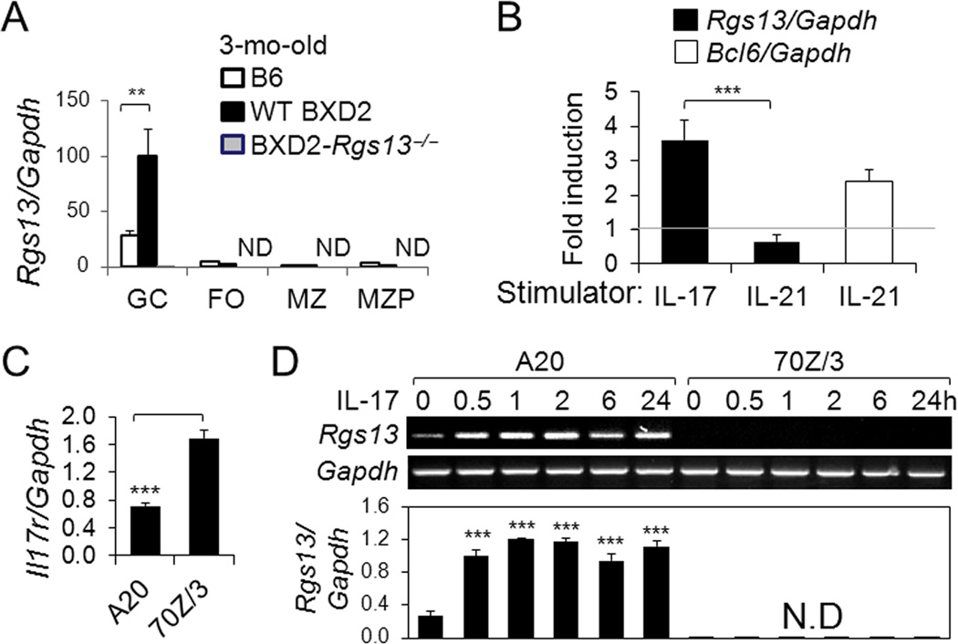
Methods: Confocal and light microscopy imaging techniques were used to determine the location of cells that express RGS13 and activation-induced cytidine deaminase (AID) in the mouse spleen, and the number of plasmablasts. The levels of GC and plasma cell program transcripts in GC B cells were determined by real-time quantitative polymerase chain reaction (qPCR). Differential interleukin-17 (IL-17)-mediated expression of RGS13 in GC versus non-GC B cells was analyzed using A20 and 70Z/3 B cells.

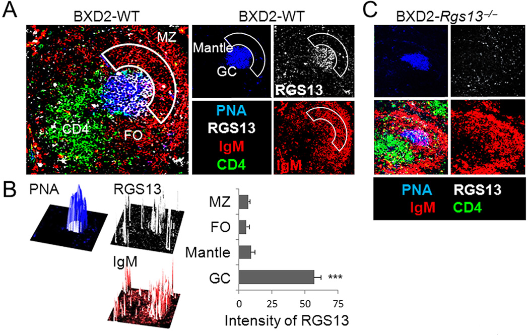
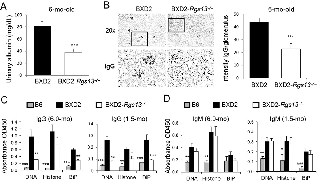
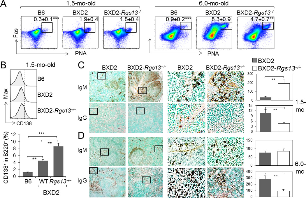
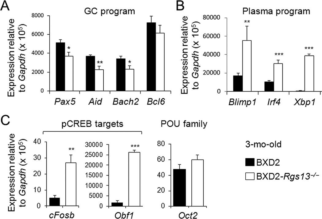
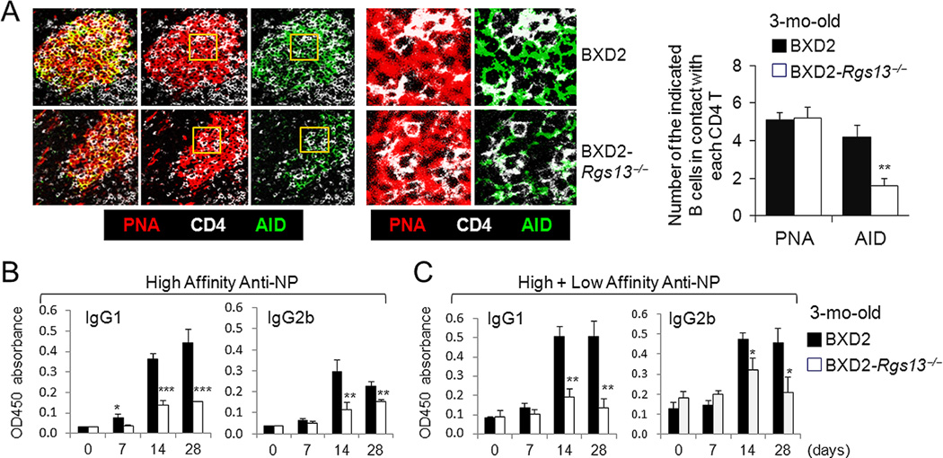
Results: In the spleens of BXD2 mice, RGS13 was mainly expressed by GC B cells and was stimulated by IL-17 but not IL-21. IL-17 up-regulated RGS13 in A20 GC cells but not 70Z/3 non-GC B cells. BXD2- Rgs13-/- mice exhibited smaller GCs and lower AID levels, suggesting lower somatic hypermutation and affinity maturation. However, GC B cells from BXD2- Rgs13-/- mice showed increased levels of IgMbright plasmablasts, up-regulation of the genes encoding plasma program, including interferon regulatory factor 4, B lymphocyte-induced maturation protein 1, and X-box binding protein 1 and the p-CREB target genes Fosb and Obf1, and down-regulation of the GC program genes Aid, Pax5, and Bach2 compared to BXD2 mice. BXD2-Rgs13-/- mice had lower titers of IgG autoantibodies and IgG deposits in the glomeruli, suggesting reduced autoantibody pathogenicity.

Conclusion: RGS13 deficiency is associated with a reduction in GC program genes and the exit of fewer pathogenic IgM plasmablasts in BXD2 mice. Our findings indicate that prolonged GC program, mediated by up-regulation of RGS13, enhances AID expression and enables the generation of pathogenic autoantibodies in autoreactive GCs.


Conflict of interest statement

All authors claim to have no financial interests which could create a potential conflict of interest or the appearance of a conflict of interest with regard to the work.

Figures

Figure 1
Figure 1
RGS13 is exclusively expressed by GC B cells. Confocal imaging photomicrographs of RGS13 expression (white) in the spleen of 3-mo-old (A, B) BXD2 or (C) control BXD2-Rgs13−/− mice. A, C, Frozen spleen sections were costained with PNA (blue), anti-IgM (red) and anti-CD4 (green)(Objective lens = 20×). Abbreviations used: MZ, marginal zone; FO, follicular area. Image-J analysis of the RGS13 color intensity relative to the color intensity of PNA and IgM by overlay of separate color projections. B, The individual 3D plots represent the section of a representative BXD2 spleen tissue examined by confocal microscopy, with the z-axis representing intensity. Intensity of RGS13 in specified areas is shown as the mean ± SEM. Four sections were evaluated with 3–5 randomly chosen follicles analyzed per section (*** p<0.005 between RGS13 in GC compared to RGS13 in other regions).
Figure 2
Figure 2
Induction of Rgs13 in GC B cells by IL-17. A, qRT-PCR analysis of Rgs13 expression in B cells sorted from the spleens of indicated strains (ND = not detectable; ** p<0.01 for the indicated comparisons). B, qRT-PCR analysis of Rgs13 after normalization to Gapdh, in purified BXD2 B cells 2 hrs after treatment with IL-17 or IL-21 (50 ng/ml) and Bcl6, after normalization to Gapdh, in purified BXD2 B cells 2 hrs after treatment with IL-21 (50 ng/ml)(Rt bar). Fold changes of the indicated genes relative to the untreated control (indicated by a gray line) are shown. C, qRT-PCR analysis of basal levels of Il17ra, after normalization to Gapdh, in A20 and 70Z/3 cells. (D) Gel electrophoresis (upper) and qRT-PCR analysis (lower) of Rgs13, after normalization to Gapdh, by A20 and 70Z/3 cells. Cells were stimulation with IL-17 at the indicated time points. Results are shown as the mean ± SEM (ND = not detectable; *** p< 0.005 between the indicated comparison or between basal versus stimulated cells)(N=4 for all panels).
Figure 3
Figure 3
Kidney disease and production of pathogenic IgG autoantibodies are reduced in BXD2-Rgs13−/− mice. A, ELISA analysis of urinary albumin in 6-mo-old mice (N=6, ***p<0.005). B, Left: Representative photomicrographs of immunohistochemical staining of IgG antibody deposits on glomeruli. Objective lens was 20×. Box indicates area that was enlarged and shown in lower panels. Right: ImageJ quantitation of the intensity of IgG deposits in glomeruli of the indicated strain at 6-mo of age. Data are shown as mean ± SEM. At least 4 kidney sections were examined per mouse, and staining of at least 10 glomeruli quantified for each kidney section. C, D, ELISA analysis of serum levels of (C) IgG and (D) IgM autoantibodies in the indicated strains at 6.0-mo and 1.5-mo of age (mean ± SEM, N=6–10 mice/group; * p<0.05, ** p<0.01, *** p<0.005 for results for each strain in comparison with age-matched BXD2-WT mice).
Figure 4
Figure 4
A deficiency of RGS13 in BXD2 mice obstructs age-related increase of GCs but enhances the numbers of IgM plasma cells in young mice. A, FACS analysis of the percent of PNA+Fas+ CD19+ GC B cells in the spleens of 1.5- and 6-mo-old mice (mean ± SEM of the percent of gated population within B cells, N=4; ** p<0.01, *** p<0.005 for the indicated strain in comparison with BXD2-WT mice). B, FACS analysis of the percent of CD138+ (Syndecan-1) B220+ plasmablasts in B6, BXD2, and BXD2-Rgs13−/− mice, at 1.5-mo-old. C, D, Left, Low power (10× objective lens) and Middle, zoomed view: Representative photomicrographs of immunohistochemical staining of IgMbright and IgGbright cells in the spleens of (C) 1.5-mo-old and (D) 6-mo-old mice. Right, Quantitation of the number of IgMbright and IgGbright cells per 10× objective lens microscopic view (mean ± SEM. N=4 mice per group). At least 3 randomly chosen areas were counted per section (** p<0.01 or *** p<0.005 between the indicated comparisons, for Panels B - D).
Figure 5
Figure 5
Decreased GC program and increased plasma program gene expression in the absence of RGS13. qRT-PCR analysis of (A) GC program-related, (B) Plasma program-related, and (C) pCREB targeted or POU family transcription factor genes (CD19+PNA+Fas+) sorted from the spleens of mice (N=2 mice per group with 3 independent measurements. * p<0.05, ** p<0.01, *** p<0.005 between BXD2 and BXD2-Rgs13−/−).
Figure 6
Figure 6
A deficiency of RGS13 in BXD2 mice limits AID expression and antibody affinity maturation. A, Confocal immunofluorescent assessment of position of PNA+ or AID+ B cells and CD4 T cells in the GCs. Yellow rectangle gated areas were enlarged to reveal the contact between CD4 T cells (white) and PNA+ GC B cells (red) or between CD4 T cells (white) and AID+ B cells (green). Bar graph showing the average numbers of PNA+ GC B cells or AID+ GC B cells surrounding each CD4 T cell in the GCs. At least 3 GCs were analyzed per section with 4 sections per strain (** p<0.01, as compared to BXD2-WT mice). B, C, ELISA analysis of the serum (B) high affinity anti-NP and (C) high+low affinity anti-NP IgG1 and IgG2b antibody responses at the indicated days after immunization of mice with NP21-CGG (N=4 mice per group; * p<0.05; ** p<0.01; *** p<0.005 as compared to BXD2-WT mice). All Data are shown as mean ± SEM.

References

    1. Schroeder K, Herrmann M, Winkler TH. The role of somatic hypermutation in the generation of pathogenic antibodies in SLE. Autoimmunity. 2012 - PubMed
    1. Holers VM. Are anti-cyclic citrullinated peptide antibodies pathogenic in rheumatoid arthritis? Nat Clin Pract Rheumatol. 2006;2(8):400–401. - PubMed
    1. Snir O, Widhe M, Hermansson M, von Spee C, Lindberg J, Hensen S, et al. Antibodies to several citrullinated antigens are enriched in the joints of rheumatoid arthritis patients. Arthritis Rheum. 2010;62(1):44–52. - PubMed
    1. Bootsma H, Spronk PE, Ter Borg EJ, Hummel EJ, de Boer G, Limburg PC, et al. The predictive value of fluctuations in IgM and IgG class anti-dsDNA antibodies for relapses in systemic lupus erythematosus. A prospective long-term observation. Ann Rheum Dis. 1997;56(11):661–666. - PMC - PubMed
    1. Arbuckle MR, McClain MT, Rubertone MV, Scofield RH, Dennis GJ, James JA, et al. Development of autoantibodies before the clinical onset of systemic lupus erythematosus. N Engl J Med. 2003;349(16):1526–1533. - PubMed

Publication types

MeSH terms