. 2014 Jan 1;522(1):169-85.
doi: 10.1002/cne.23394.

Distribution of language-related Cntnap2 protein in neural circuits critical for vocal learning

Affiliations

Distribution of language-related Cntnap2 protein in neural circuits critical for vocal learning

Michael C Condro et al. J Comp Neurol. .

Abstract

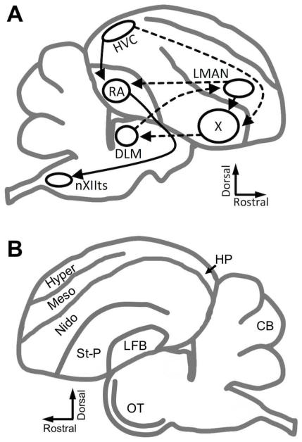
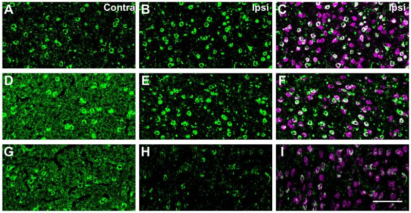
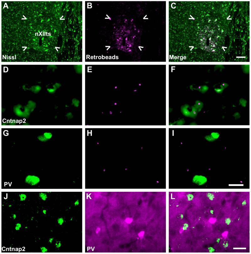
Variants of the contactin associated protein-like 2 (Cntnap2) gene are risk factors for language-related disorders including autism spectrum disorder, specific language impairment, and stuttering. Songbirds are useful models for study of human speech disorders due to their shared capacity for vocal learning, which relies on similar cortico-basal ganglia circuitry and genetic factors. Here we investigate Cntnap2 protein expression in the brain of the zebra finch, a songbird species in which males, but not females, learn their courtship songs. We hypothesize that Cntnap2 has overlapping functions in vocal learning species, and expect to find protein expression in song-related areas of the zebra finch brain. We further expect that the distribution of this membrane-bound protein may not completely mirror its mRNA distribution due to the distinct subcellular localization of the two molecular species. We find that Cntnap2 protein is enriched in several song control regions relative to surrounding tissues, particularly within the adult male, but not female, robust nucleus of the arcopallium (RA), a cortical song control region analogous to human layer 5 primary motor cortex. The onset of this sexually dimorphic expression coincides with the onset of sensorimotor learning in developing males. Enrichment in male RA appears due to expression in projection neurons within the nucleus, as well as to additional expression in nerve terminals of cortical projections to RA from the lateral magnocellular nucleus of the nidopallium. Cntnap2 protein expression in zebra finch brain supports the hypothesis that this molecule affects neural connectivity critical for vocal learning across taxonomic classes.

Keywords: Caspr2; autism; birdsong; speech; zebra finch.


Figures

Figure 1
Figure 1. Diagram of the songbird brain
A) Schematic sagittal drawing depicts simplified song control circuitry. Solid lines indicate the posterior motor pathway, beginning with HVC, which projects to RA. RA directly projects to nXIIts, which controls the motor neurons of the syrinx. Dashed lines indicate connections of the AFP, in which HVC, X, DLM, and LMAN comprise a cortical-basal ganglia-thalamo-cortical loop like those underlying procedural learning in mammalian brains. LMAN completes the song circuit by projecting to RA, as well as back to X. B) Schematic sagittal drawing depicts non-song brain regions in which Cntnap2 immunostaining was analyzed in this study.
Figure 2
Figure 2. Antibody detection of zebra finch Cntnap2
A) Western blot of zebra finch whole brain homogenate. Anti-Cntnap2 primary antibody detects a single prominent protein band at the predicted molecular weight (~180 kDa) for endogenous zebra finch Cntnap2. B) Western blots of the ZFTMA zebra finch established cell line with a plasmid expressing zebra finch Cntnap2 or GFP. Transfection of the Cntnap2 construct results in a detectable signal at the predicted molecular weight for Cntnap2 (left). In contrast, transfection of GFP results in no detectable signal at the same molecular weight, confirming no endogenous Cntnap2 expression in this skin-derived cell line (right). For each condition, preadsorption of the primary antibody with its antigenic peptide (Ab+pep) dramatically reduces or removes the signal. Molecular weight markers are given in kDa. C-E) Zebra finch optic and F-H) sciatic nerves double labeled with Cntnap2 and potassium channel subunit Kvβ2 antibodies. Cntnap2 signal colocalizes with putative signals for potassium channel subunit Kvβ2 in both nerves, consistent with its expression in rodents (Poliak et al., 1999; 2003). Overlap of these signals in zebra finch nerves further validates the Cntnap2 antibody for use in this model. Scale bars = 10 μm C-E; 5 μm F-H.
Figure 3
Figure 3. Cntnap2 distribution in non-song circuit brain regions
Cntnap2 is detected in several areas outside the song circuit of the zebra finch brain, including in structures reported to express Cntnap2 in rodents (Poliak et al., 1999). Non-song circuit tissue in this figure are taken from regions depicted in Fig. 1B. Neuron specific marker NeuN (magenta) is used for reference. A-C) Axonal patterning of Cntnap2 label in the lateral forebrain bundle within the telencephalon. D-F) intense Cntnap2 (green) labeling along axons in layer 5 of the optic tectum. Numbers in 3B indicate the layers of the optic tectum according to Ramón y Cajal (1911). G-I) Purkinje cell bodies and the cerebellar white matter strongly express Cntnap2, with less in the molecular layer, and fibrous signal in the granular layer and white matter. J-L) Coronal section of the medial hippocampus; numbers indicate layers (Montagnese et al., 1996). Cntnap2 marks neuronal somata in the pyramidal cell region (white arrows). Scale bars = 50 μm A-C; 200 μm D-L.
Figure 4
Figure 4. Cntnap2 protein in song circuit nuclei
Fluorescent photomicrographic images of song control nuclei. Cntnap2 signals are in green, and NeuN signals in magenta. A-C) HVC in the nidopallium; D-F) RA in the arcopallium; G-I) LMAN in the nidopallium; J-L) Area X in the striatopallidum, inset: higher magnification inside X; M-O) DLM in the thalamus, along with the ovoid nucleus, the dorsomedial nucleus of the posterior thalamus, and the lateral forebrain bundle. Each nucleus is indicated by dashed line traces on the NeuN (middle column) panels. Greater Cntnap2 labeling is found within RA, LMAN, and area X relative to surrounding brain regions on both cell bodies and in the neuropil. HVC and DLM contain Cntnap2-expressing cells, but with expression levels comparable to their surrounding tissues. Scale bars = 200 μm A-I; 100 μm J-L (50 μm inset); 200 μm M-O.
Figure 5
Figure 5. Cntnap2 within RA in both sexes at developmental time points during male song learning
A-J) Representative images of Cntnap2 immunolabeling of cells in male (A-E) and female (F-J) RA at time points during development encompassing the onset of sensory acquisition, sensorimotor learning, and crystallization of song. Anti-NeuN signals (not shown) were used to trace the border of RA in each image. As previously reported (Konishi and Akutagawa, 1985; Nixdorf-Bergweiler, 1996), the size of RA begins to decrease in females and increase in males starting around 35d and continues through development until maturity. K) A diagram of RA and the two arcopallial regions in which labeled cells were counted: the ventral intermediate arcopallium (AIV) and the dorsal arcopallium (AD). L-N) Graphs representing the percentage of Cntnap2 positive neurons out of the total number of NeuN positive cells found in RA, AIV, and AD, respectively, for 3-6 birds of each sex at each time point. Statistical significance was determined by resampling ANOVA, followed by individual Student’s T-tests *p<0.05, **p<0.01. Scale bar = 100 μm.
Figure 6
Figure 6. Unilateral LMAN lesions result in an ipsilateral decrease of Cntnap2 in RA
Representative photomicrographic images of Cntnap2 labeling (green) in RA from three adult male zebra finches (A-C, D-F, G-I) in which LMAN was lesioned unilaterally by injection of ibotenic acid. Double labeling with NeuN (magenta; C,F,I) indicates neuronal cell bodies. In all cases, the lesion reduces the amount of Cntnap2 in the neuropil, but not cell bodies, in ipsilateral RA relative to the contralateral nucleus, indicating that some of the Cntnap2 in the neuropil originates from LMAN projections. Scale bar = 25 μm.
Figure 7
Figure 7. Cntnap2 is expressed in RA projection neurons, not parvalbumin positive interneurons
A-C) Injection site of retrobeads (magenta) in nXIIts (indicated by white arrows), identified by Nissl stain (green). D-F) Retrobeads overlap with Cntnap2 (green) expressing cells in RA. G-I). Retrobeads do not overlap with strongly parvalbumin positive interneurons. J-L) Cntnap2 immunolabeling (green) does not overlap with RA inhibitory interneurons intensely labeled with parvalbumin (magenta). Retrograde labeling reveals that RA projection neurons express Cntnap2 and confirms its absence in parvalbumin positive interneurons. Scale bar = 50 μm A-C; 20 μm D-I; 25 μm J-L.

References

    1. Abrahams BS, Tentler D, Perederiy JV, Oldham MC, Coppola G, Geschwind DH. Genome-wide analyses of human perisylvian cerebral cortical patterning. Proc Natl Acad Sci. 2007;104:17849–17854. - PMC - PubMed
    1. Alarcón M, Abrahams BS, Stone JL, Duvall JA, Perederiy JV, Bomar JM, Sebat J, Wigler M, Martin CL, Ledbetter DH, et al. Linkage, association, and gene-expression analyses identify CNTNAP2 as an autism-susceptibility gene. Am J Hum Genet. 2008;82:150–159. - PMC - PubMed
    1. Anderson GR, Galfin T, Xu W, Aoto J, Malenka RC, Südhof TC. Candidate autism gene screen identifies critical role for cell-adhesion molecule CASPR2 in dendritic arborization and spine development. Proc Natl Acad Sci. 2012;109(44):18120–5. - PMC - PubMed
    1. Arking DE, Cutler DJ, Brune CW, Teslovich TM, West K, Ikeda M, Rea A, Guy M, Lin S, Cook EH, et al. A common genetic variant in the neurexin superfamily member CNTNAP2 increases familial risk of autism. Am J Hum Genet. 2008;82:160–164. - PMC - PubMed
    1. Arnold AP, Chen X, Link JC, Itoh Y, Reue K. Cell-autonomous sex determination outside of the gonad. Dev Dyn. 2013;242:371–379. - PMC - PubMed

Publication types