Lipid in skeletal muscle myotubes is associated to the donors' insulin sensitivity and physical activity phenotypes
- PMID: 23818429
- PMCID: PMC3883809
- DOI: 10.1002/oby.20556
Lipid in skeletal muscle myotubes is associated to the donors' insulin sensitivity and physical activity phenotypes
Abstract
Objective: This study investigated the relationship between in vitro lipid content in myotubes and in vivo whole body phenotypes of the donors such as insulin sensitivity, intramyocellular lipids (IMCL), physical activity, and oxidative capacity.
Design and methods: Six physically active donors were compared to six sedentary lean and six T2DM. Lipid content was measured in tissues and myotubes by immunohistochemistry. Ceramides, triacylglycerols, and diacylglycerols (DAGs) were measured by LC-MS-MS and GC-FID. Insulin sensitivity was measured by hyperinsulinemic-euglycemic clamp (80 mU min⁻¹ m⁻²), maximal mitochondrial capacity (ATPmax) by ³¹P-MRS, physical fitness by VO₂max and physical activity level (PAL) by accelerometers.
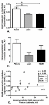
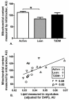
Results: Myotubes cultured from physically active donors had higher lipid content (0.047 ± 0.003 vs. 0.032 ± 0.001 and 0.033 ± 0.001AU; P < 0.001) than myotubes from lean and T2DM donors. Lipid content in myotubes was not associated with IMCL in muscle tissue but importantly, correlated with in vivo measures of ATPmax (r = 0.74; P < 0.001), insulin sensitivity (r = 0.54; P < 0.05), type-I fibers (r = 0.50; P < 0.05), and PAL (r = 0.92; P < 0.0001). DAGs and ceramides in myotubes were inversely associated with insulin sensitivity (r = -0.55, r = -0.73; P < 0.05) and ATPmax (r = -0.74, r = -0.85; P < 0.01).
Conclusions: These results indicate that cultured human myotubes can be used in mechanistic studies to study the in vitro impact of interventions on phenotypes such as mitochondrial capacity, insulin sensitivity, and physical activity.
Trial registration: ClinicalTrials.gov NCT00401791 NCT00402012.
Copyright © 2013 The Obesity Society.
Figures


References
-
- Pan DA, Lillioja S, Kriketos AD, et al. Skeletal muscle triglyceride levels are inversely related to insulin action. Diabetes. 1997;46:983–8. - PubMed
-
- Goodpaster BH, He J, Watkins S, Kelley DE. Skeletal muscle lipid content and insulin resistance: evidence for a paradox in endurance-trained athletes. J Clin Endocrinol Metab. 2001;86:5755–61. - PubMed
-
- Moro C, Bajpeyi S, Smith SR. Determinants of intramyocellular triglyceride turnover: implications for insulin sensitivity. Am J Physiol Endocrinol Metab. 2008;294:E203–13. - PubMed
Publication types
MeSH terms
Substances
Associated data
Grants and funding
LinkOut - more resources
Full Text Sources
Other Literature Sources
Medical
Research Materials
Miscellaneous

