. 2013 Jul 23;110(30):12480-5.
doi: 10.1073/pnas.1310261110. Epub 2013 Jul 1.

Fat cells directly sense temperature to activate thermogenesis

Affiliations

Fat cells directly sense temperature to activate thermogenesis

Li Ye et al. Proc Natl Acad Sci U S A. .

Abstract

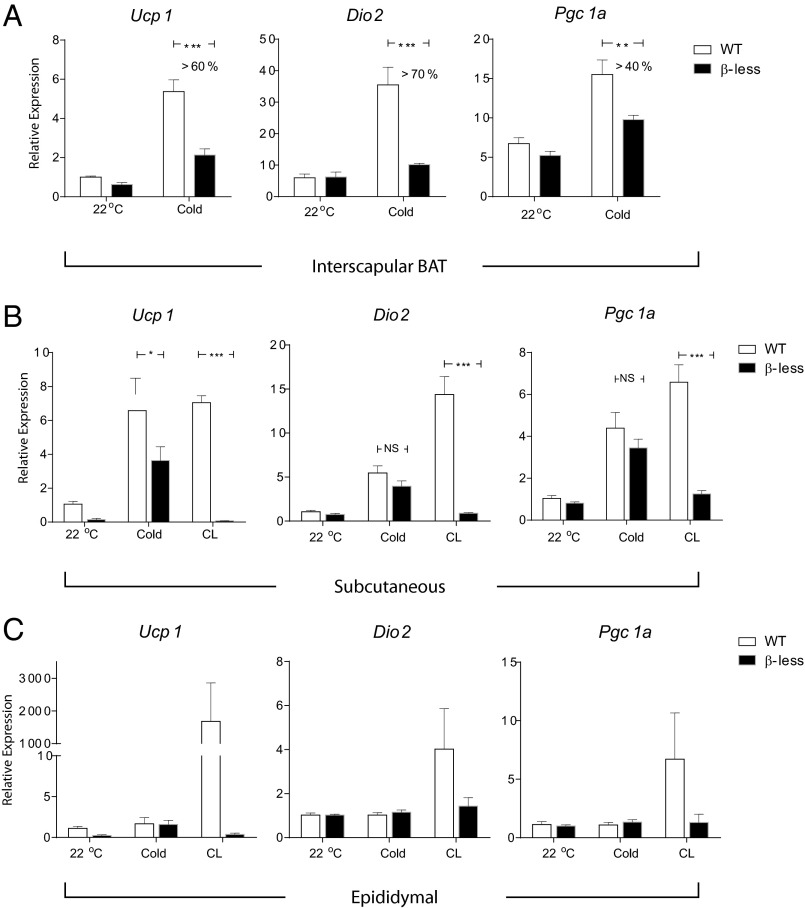
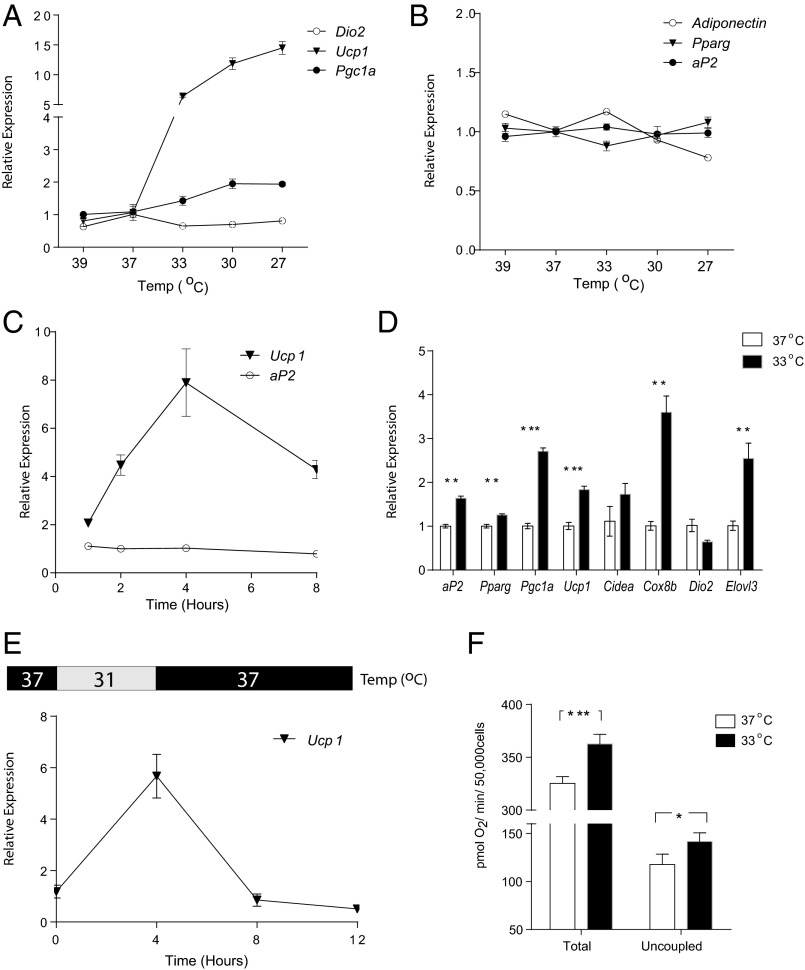
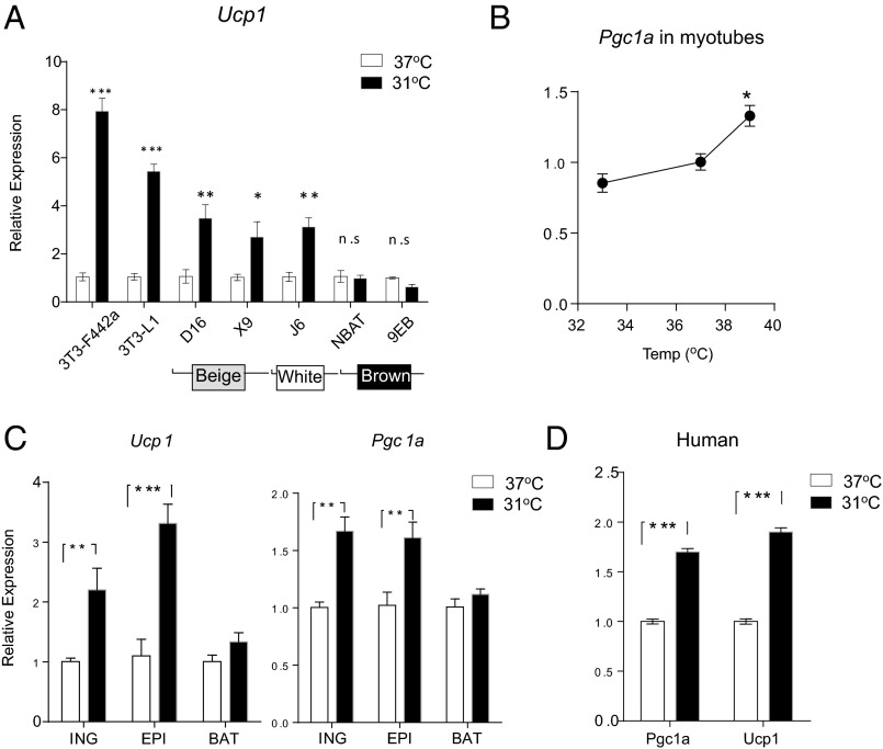
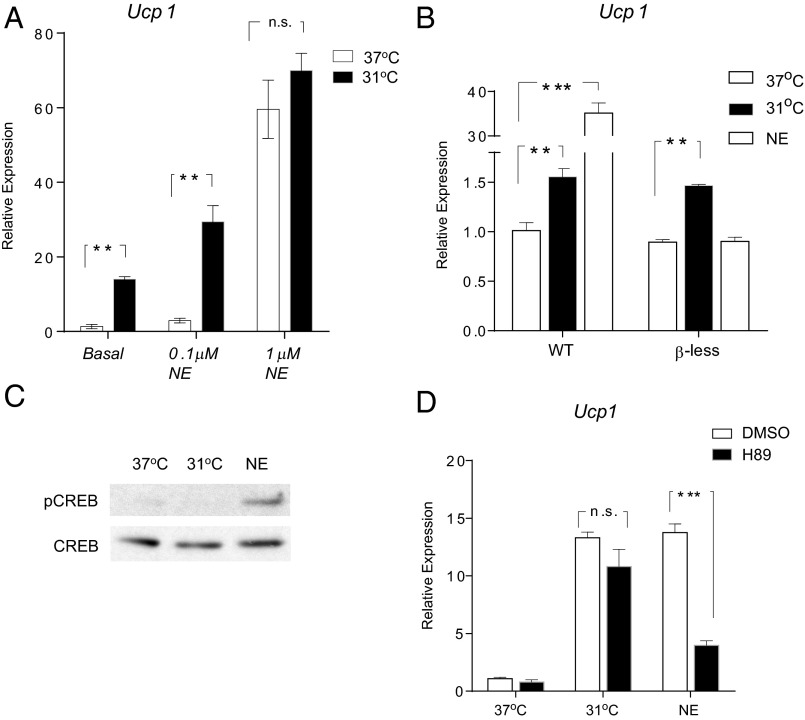
Classic brown fat and inducible beige fat both dissipate chemical energy in the form of heat through the actions of mitochondrial uncoupling protein 1. This nonshivering thermogenesis is crucial for mammals as a defense against cold and obesity/diabetes. Cold is known to act indirectly through the sympathetic nervous systems and β-adrenergic signaling, but here we report that cool temperature (27-33 °C) can directly activate a thermogenic gene program in adipocytes in a cell-autonomous manner. White and beige fat cells respond to cool temperatures, but classic brown fat cells do not. Importantly, this activation in isolated cells is independent of the canonical cAMP/Protein Kinase A/cAMP response element-binding protein pathway downstream of the β-adrenergic receptors. These findings provide an unusual insight into the role of adipose tissues in thermoregulation, as well as an alternative way to target nonshivering thermogenesis for treatment of obesity and metabolic diseases.

Keywords: Ucp1; cold sensing.


Conflict of interest statement

The authors declare no conflict of interest.

Figures

Fig. 1.
Fig. 1.
Cold induces a β-adrenergic–independent thermogenic gene program in s.c. adipose tissue. qPCR analysis of Ucp1, Dio2, and Pgc1a mRNA in interscapular brown fat (A), s.c. fat (inguinal) (B), and visceral fat (epididymal) (C) from β-less and WT mice. For cold exposure, mice were individually housed in 10 °C for 20 h. As a positive control, 24-h treatment of CL316243 (1 mg/kg, twice daily) was given. Data are presented as mean ± SEM (n = 6–8 in each group).
Fig. 2.
Fig. 2.
Cool temperatures induce a thermogenic program in isolated white adipocytes. Cultured and fully differentiated 3T3-F442A adipocytes were exposed to 39 °C, 37 °C, 33 °C, 30 °C, and 27 °C for 4 h before mRNA was harvested (Materials and Methods). The mRNA expression of thermogenic genes (A) and adipose markers (B) were measured by qPCR. (C) 3T3-F442A adipocytes were exposed to 31 °C for 1, 2, 4, and 8 h before mRNA was harvested and analyzed by qPCR. The values were normalized to cells kept in 37 °C from the same times. (D) 3T3-F442A adipocytes were differentiated and maintained in 37 °C or 33 °C for 10 d (day 0–10). mRNA was analyzed by qPCR for thermogenic gene expression. (E) 3T3-F442A adipocytes were first exposed to 31 °C for 4 h, and then the temperature was changed back to 37 °C for an additional 4–8 h. mRNA was analyzed by qPCR and normalized to cells kept in 37 °C from the same times. (F) Total and uncoupled respiration (as oxygen consumption rate) in 3T3-F442A adipocytes were measured after 6 h incubation in 37 °C or 31 °C. Data are presented as mean ± SD (n = 6 in each group).
Fig. 3.
Fig. 3.
The temperature-induced thermogenic program is specific to white and beige adipocytes. (A) Multiple fat cell lines were exposed to 31 °C for 4 h before Ucp1 mRNA expression was measured by qPCR. (B) Differentiated C2C12 myotubes were exposed to 33 °C, 37 °C, or 39 °C for 4 h. mRNA was harvested and analyzed by qPCR for Pgc1a expression. Primary mouse adipocytes from inguinal, epididymal, and interscapular brown fat (C) and primary human adipocytes from s.c. fat (D) were exposed to 31 °C for 4 h. mRNA expression of Ucp1 and Pgc1a was measured by qPCR. Data are presented as mean ± SD (n = 6 in each group).
Fig. 4.
Fig. 4.
The temperature-induced thermogenic program is independent of the canonical pathway. (A) Fully differentiated 3T3-F442A adipocytes were exposed to PBS (as “Basal”) or NE at indicated doses for 4 h at 37 °C or 31 °C. Ucp1 mRNA was measured by qPCR. (B) WT and β-less adipocytes (derived from the inguinal depots) were exposed to 31 °C or treated with 100 nM NE for 4 h before Ucp1 mRNA was measured by qPCR. (C) Fully differentiated 3T3-F442A adipocytes were exposed to 31 °C or 100 nM NE for 20 min, and cell lysates were analyzed by Western blot. (D) 3T3-F442A adipocytes were exposed to 31 °C or 100 nM NE for 4 h, with a 30-min pretreatment of either 10 μM H89 or DMSO. Ucp1 mRNA was analyzed by qPCR. Data are presented as mean ± SD (n = 6 in each group).

References

    1. Cannon B, Nedergaard J. Brown adipose tissue: Function and physiological significance. Physiol Rev. 2004;84(1):277–359. - PubMed
    1. Lowell BB, et al. Development of obesity in transgenic mice after genetic ablation of brown adipose tissue. Nature. 1993;366(6457):740–742. - PubMed
    1. Feldmann HM, Golozoubova V, Cannon B, Nedergaard J. UCP1 ablation induces obesity and abolishes diet-induced thermogenesis in mice exempt from thermal stress by living at thermoneutrality. Cell Metab. 2009;9(2):203–209. - PubMed
    1. Cypess AM, et al. Identification and importance of brown adipose tissue in adult humans. N Engl J Med. 2009;360(15):1509–1517. - PMC - PubMed
    1. van Marken Lichtenbelt WD, et al. Cold-activated brown adipose tissue in healthy men. N Engl J Med. 2009;360(15):1500–1508. - PubMed

Publication types

Substances