. 2013 Jul 16;110(29):11827-32.
doi: 10.1073/pnas.1300325110. Epub 2013 Jul 1.

Posttranslational modification of CENP-A influences the conformation of centromeric chromatin

Affiliations

Posttranslational modification of CENP-A influences the conformation of centromeric chromatin

Aaron O Bailey et al. Proc Natl Acad Sci U S A. .

Abstract

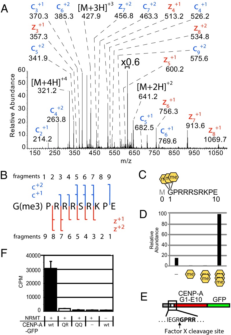
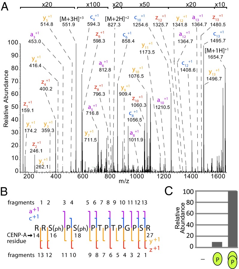
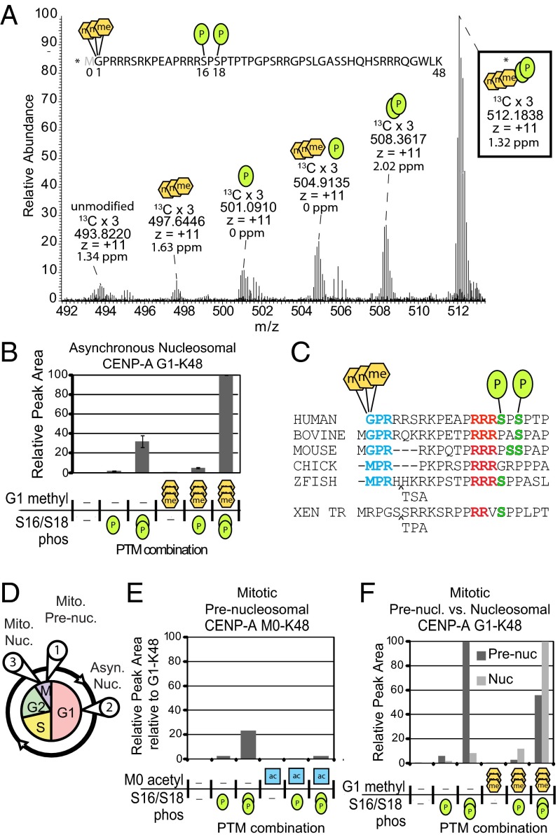
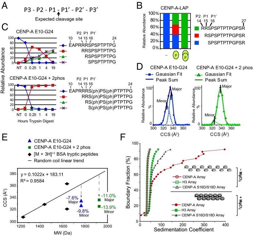
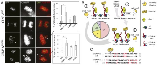
Centromeres are chromosomal loci required for accurate segregation of sister chromatids during mitosis. The location of the centromere on the chromosome is not dependent on DNA sequence, but rather it is epigenetically specified by the histone H3 variant centromere protein A (CENP-A). The N-terminal tail of CENP-A is highly divergent from other H3 variants. Canonical histone N termini are hotspots of conserved posttranslational modification; however, no broadly conserved modifications of the vertebrate CENP-A tail have been previously observed. Here, we report three posttranslational modifications on human CENP-A N termini using high-resolution MS: trimethylation of Gly1 and phosphorylation of Ser16 and Ser18. Our results demonstrate that CENP-A is subjected to constitutive initiating methionine removal, similar to other H3 variants. The nascent N-terminal residue Gly1 becomes trimethylated on the α-amino group. We demonstrate that the N-terminal RCC1 methyltransferase is capable of modifying the CENP-A N terminus. Methylation occurs in the prenucleosomal form and marks the majority of CENP-A nucleosomes. Serine 16 and 18 become phosphorylated in prenucleosomal CENP-A and are phosphorylated on asynchronous and mitotic nucleosomal CENP-A and are important for chromosome segregation during mitosis. The double phosphorylation motif forms a salt-bridged secondary structure and causes CENP-A N-terminal tails to form intramolecular associations. Analytical ultracentrifugation of phospho-mimetic CENP-A nucleosome arrays demonstrates that phosphorylation results in greater intranucleosome associations and counteracts the hyperoligomerized state exhibited by unmodified CENP-A nucleosome arrays. Our studies have revealed that the major modifications on the N-terminal tail of CENP-A alter the physical properties of the chromatin fiber at the centromere.

Keywords: epigenetics; kinetochore; mass spectrometry.


Conflict of interest statement

The authors declare no conflict of interest.

Figures

Fig. 1.
Fig. 1.
CENP-A is trimethylated on the α-amino position of the N-terminal glycine. (A) CENP-A GluC digestions produced an ETD MS2 spectrum of an N-terminal peptide trimethylated on the α-N position of glycine 1. Value above bracket indicates magnification. (B) Sequence coverage of α-N trimethylated CENP-A ETD MS2 spectrum. (C) GluC cleavages at E10 to produce an N-terminal peptide that had been subjected to proteolytic initiating methionine removal in vivo. A conventional histone nomenclature is adopted for CENP-A where the initiator methionine is “Met0.” (D) Integrated chromatographic peak areas of CENP-A G1–E10 methylated forms. Orange hexagons, α-N methylation. (E) A fusion protein containing amino acids 1–10 of CENP-A was engineered to reveal Gly1 by Factor X cleavage to produce a substrate for NRMT modification. (F) CENP-A fusion protein was methylated using recombinant NRMT and 3H-SAM. Wild-type CENP-A N termini (GPRR-) and mutants in which R3 and R4 were replaced by glutamine (GPQR- or GPQQ-) were tested. P < 0.01, Student’s t test, n = 3 independent experiments ± SD.
Fig. 2.
Fig. 2.
The CENP-A N-terminal tail is doubly phosphorylated at Ser16 and Ser18. (A) ETD MS2 spectrum of doubly phosphorylated CENP-A R14–R27. Value at bracket indicates magnification level for region of spectrum. (B) Sequence coverage of an MS2 spectrum of a trypsin-generated CENP-A peptide R14–R27. Signature a, c, y, and z ions indicate the presence of phosphate at Ser16 and Ser18. (C) CENP-A is unmodified and singly or doubly phosphorylated in a ratio of ∼1:10:100, respectively. Green ovals represent degree of phosphorylation on peptide.
Fig. 3.
Fig. 3.
CENP-A N-terminal tails are marked with combinations of stable phosphorylation and α-N methylation. (A) LysC digestion yields a CENP-A G1–K48 peptide. A full MS spectrum (17 averaged scans) shows combinatorial α-N trimethylation and phosphorylations of the same peptides in asynchronously cycling cells. Accurate mass and charge state is reported for the 13C x 3 isotopes. The most abundant form is trimethylated at Gly1 and doubly phosphorylated at Ser16 and Ser18. Green oval, phosphorylation; orange hexagon, methylation. (B) Integrated chromatographic peak areas of CENP-A G1–K48 PTM forms. n = 3 independent biological replicates. Error bars represent SEM. (C) Comparison of vertebrate CENP-A protein sequences reveals conservation at observed PTM sites: N termini (blue), proline–arginine (red), and serine (green). (D) Soluble and chromatin fractions of cells blocked in mitosis were used to purify populations of (circle 1) prenucleosomal, and nucleosomal CENP-A from (circle 2) asynchronously-dividing cells and (circle 3) mitotic cells. Integrated chromatographic peak area quantified CENP-A tail PTM forms: (E) prenucleosomal containing Metinit; (F) prenucleosomal and mitotic nucleosomal fractions lacking Metinit. Acetylation (blue square) was observed only when Metinit was present.
Fig. 4.
Fig. 4.
CENP-A Ser16/Ser18 phosphorylation forms a compact secondary structure. (A) Nomenclature for proteolytic sites. (B) The percentage of trypsin cleavage at the P1–P1′, P1–P2, and P2-P3 sites for peptides derived from affinity-purified CENP-A. (C) The percentage of trypsin cleavage at the P1–P1′, P1–P2, and P2–P3 sites for unmodified and phosphorylated synthetic CENP-A peptides E10–G24 digested with trypsin over time were followed by liquid chromatography (LC)-MS. (D) IM-MS analysis of the monomeric [M + 3H]+3 ion of unmodified and doubly phosphorylated E10–G24 peptide is used to calculate the ion-neutral collision cross section (CCS) for each species. CCS values for the major- and minor-contributing conformers were calculated as Gaussian peaks from the sum of each peptide. (E) Tryptic digested BSA peptides analyzed by IM-MS are plotted according to CCS and molecular weight. Comparison of phosphorylated (square) and unmodified (triangle) E10–G24 peptide with the random coil trend line reveals that phosphorylation causes greater compactness relative to the increase in molecular weight. (F) AUC profiles of in vitro assembled chromatin arrays showing differential MgCl2-dependent folding between H3, CENP-A, and phospho-mimetic (S16D/S18D) CENP-A arrays. All array types (minus MgCl2) have overlapping sedimentation profiles, indicating that they are saturated to the same extent. Therefore, the observed differences upon array folding are exclusively due to the contributions of different proteins.
Fig. 5.
Fig. 5.
CENP-A Ser16/Ser18 phosphorylation is required for normal mitosis. (A) HCT116 cells transiently expressing histone H2B-mRFP and GFP-tagged wild-type or S16/S18A CENP-A mutants. The percentage of cells with unaligned metaphase (arrowhead) and lagging anaphase (arrow) chromosomes is higher in the S16/S18A mutant. *P = 0.018, **P = 0.002, Student’s t test, n = 3 independent experiments ± SD. (B) CENP-A PTM begins at translation and persists through the cell cycle. CENP-A α-N trimethylation occurs on a minority of the new prenucleosomal CENP-A and increases with cell cycle progression, constituting greater than 90% of the mitotic nucleosomes. (C) Human CENP-A PTM sites Gly1 α-N methylation and Ser16/Ser18 phosphorylation are especially divergent compared with H3.1. Conserved residues are in red.

References

    1. Stellfox ME, Bailey AO, Foltz DR. Putting CENP-A in its place. Cell Mol Life Sci. 2013;70(3):387–406. - PMC - PubMed
    1. Cleveland DW, Mao Y, Sullivan KF. Centromeres and kinetochores: From epigenetics to mitotic checkpoint signaling. Cell. 2003;112(4):407–421. - PubMed
    1. Blower MD, Sullivan BA, Karpen GH. Conserved organization of centromeric chromatin in flies and humans. Dev Cell. 2002;2(3):319–330. - PMC - PubMed
    1. Foltz DR, et al. The human CENP-A centromeric nucleosome-associated complex. Nat Cell Biol. 2006;8(5):458–469. - PubMed
    1. Okada M, et al. The CENP-H-I complex is required for the efficient incorporation of newly synthesized CENP-A into centromeres. Nat Cell Biol. 2006;8(5):446–457. - PubMed

Publication types

MeSH terms