. 2013 Sep 15;591(18):4515-33.
doi: 10.1113/jphysiol.2013.259382. Epub 2013 Jul 1.

Network interactions within the canine intrinsic cardiac nervous system: implications for reflex control of regional cardiac function

Affiliations

Network interactions within the canine intrinsic cardiac nervous system: implications for reflex control of regional cardiac function

Eric Beaumont et al. J Physiol. .

Abstract

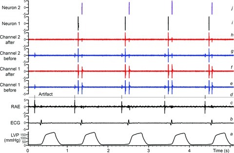
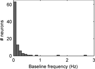
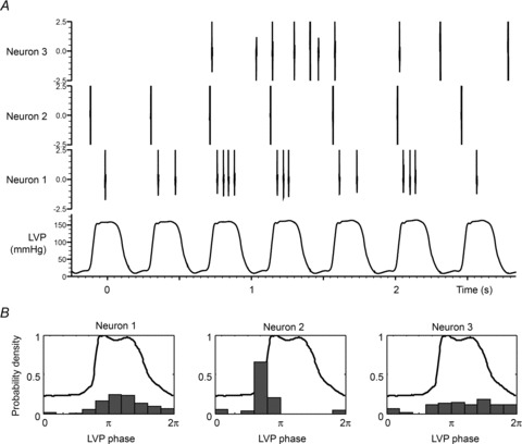
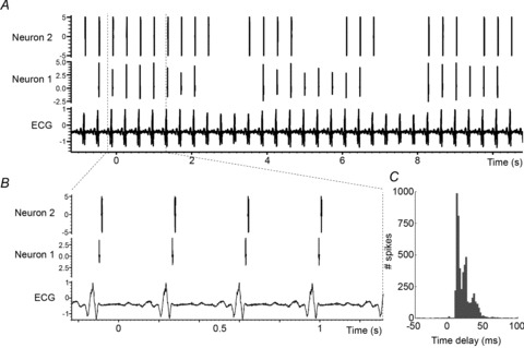
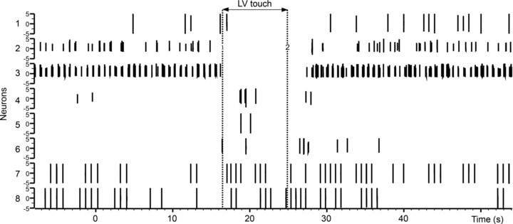
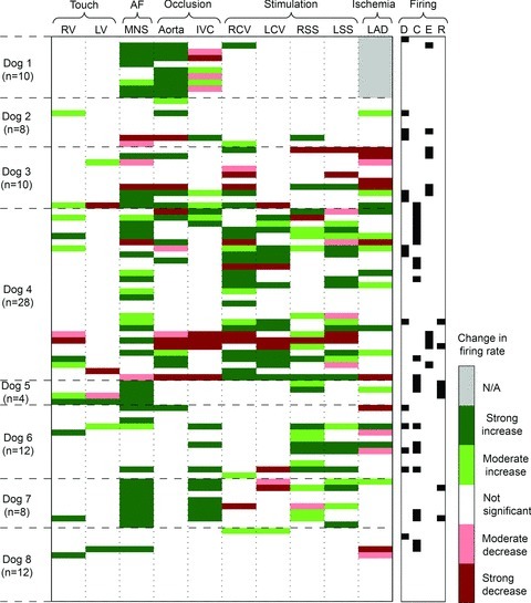
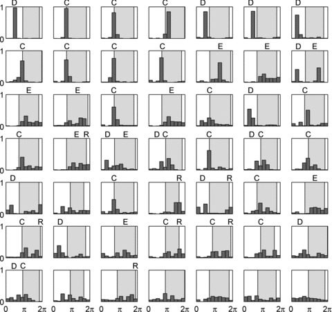
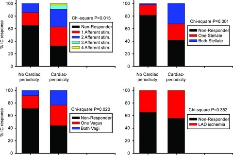
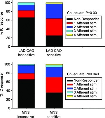
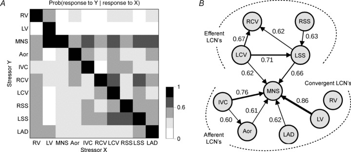
The aims of the study were to determine how aggregates of intrinsic cardiac (IC) neurons transduce the cardiovascular milieu versus responding to changes in central neuronal drive and to determine IC network interactions subsequent to induced neural imbalances in the genesis of atrial fibrillation (AF). Activity from multiple IC neurons in the right atrial ganglionated plexus was recorded in eight anaesthetized canines using a 16-channel linear microelectrode array. Induced changes in IC neuronal activity were evaluated in response to: (1) focal cardiac mechanical distortion; (2) electrical activation of cervical vagi or stellate ganglia; (3) occlusion of the inferior vena cava or thoracic aorta; (4) transient ventricular ischaemia, and (5) neurally induced AF. Low level activity (ranging from 0 to 2.7 Hz) generated by 92 neurons was identified in basal states, activities that displayed functional interconnectivity. The majority (56%) of IC neurons so identified received indirect central inputs (vagus alone: 25%; stellate ganglion alone: 27%; both: 48%). Fifty per cent transduced the cardiac milieu responding to multimodal stressors applied to the great vessels or heart. Fifty per cent of IC neurons exhibited cardiac cycle periodicity, with activity occurring primarily in late diastole into isovolumetric contraction. Cardiac-related activity in IC neurons was primarily related to direct cardiac mechano-sensory inputs and indirect autonomic efferent inputs. In response to mediastinal nerve stimulation, most IC neurons became excessively activated; such network behaviour preceded and persisted throughout AF. It was concluded that stochastic interactions occur among IC local circuit neuronal populations in the control of regional cardiac function. Modulation of IC local circuit neuronal recruitment may represent a novel approach for the treatment of cardiac disease, including atrial arrhythmias.


Figures

Figure 1
Figure 1. Methodology for the identification of individual IC neurons
Traces indicate: a, left ventricular chamber pressure (LVP); b, electrocardiogram (ECG); c, right atrial electrogram (RAE); d, event channel created from identified electrical/mechanical artifacts; e and g, raw signal recordings of two channels from the multichannel linear microarray electrode; f and h, neuronal recordings from channels e and g after artifact removal based on the event channel d; i and j, two final neuronal waveforms extracted from a stereotrode built from channels f and h using principal component analysis. These final waveforms (i and j) represent basal activity from two separate IC neurons located within the right atrial ganglionated plexus; such activity can be evaluated continuously and concurrently for hours and in response to imposed stressors.
Figure 2
Figure 2. Quantitative assessment of significance (P values) when comparing the firing rate in two intervals: baseline to stress-evoked response
P values are computed using the Skellam test and displayed as a function of the average firing rate in the first and in the second interval. The dark red and light red colours mean that the firing rate is strongly (P < 0.01) or moderately decreasing (0.01 < P < 0.05), respectively. The dark green and light green colours mean that the firing rate is strongly (P < 0.01) or moderately increasing (0.01 < P < 0.05), respectively. A, both intervals last 60 s (baseline interval 1 and response during stressor interval 2). B, the first interval (baseline) has a duration of 60 s and the second (response during stressor) has a duration of 5 s.
Figure 3
Figure 3
Histogram of baseline frequencies of all identified IC neurons
Figure 4
Figure 4. Subpopulations of IC neurons demonstrate cardiac-related neuronal activity
A, LV pressure and representative examples of neurons that are primarily active during left ventricular ejection (neuron 1), during isovolumetric contraction phase for LV (neuron 2) or with activity independent of LVP (neuron 3). B, for each of these neurons, the probability density of firing as a function of the position within the LVP cycle (expressed as a phase between 0 and 2π) is indicated along with the average LVP profile.
Figure 5
Figure 5. Long-term interdependent activity of two IC cardiovascular-related neurons
A, ECG and concurrent spontaneous activity of two IC neurons. B, zoom of A over 4 cardiac cycles. C, spike-triggered (neuron 1 to neuron 2) histogram of spontaneous activity for these two IC neurons recorded over 2 h. Note maintained temporal relationship, but with some variation in such interdependent activity.
Figure 6
Figure 6. Left ventricular touch differentially modifies IC activity
The spiking activities concurrently recorded from 8 selected IC neurons in a single animal are shown. Vertical dotted lines indicate onset and offset of touch. Note that subpopulations of IC neurons show diminished activity during touch (e.g. neurons 2 and 3), some are activated by touch (e.g. neuron 7) and some are unaffected (e.g. neuron 8).
Figure 7
Figure 7. Varied responses displayed by each neuron studied in response to differing sensory or central efferent neuronal stressors
Each horizontal column represents how each identified right atrial neuron in all 8 dogs responded to each of the stressors applied (horizontal row above). Each column is associated with a specific stressor: afferent activation (touch of right (RV) or left (LV) ventricle; occlusion of inferior vena cava (IVC) or descending aorta; myocardial ischaemia evoked by transient occlusion of left anterior descending coronary artery (LAD)), activation of efferent inputs to the RAGP via electrical stimulation of the right (RCV) or left (LCV) cervical vagus or stellate ganglia (right, RSS; left, LSS), or global activation of the IC network evoked by electrical stimulation of mediastinal nerves (MNS) at levels sufficient to evoke atrial fibrillation (AF). The number (n) of neurons so identified in each animal is indicated to left. The significance of each change ranged from greatest (P < 0.01) to moderate (0.01 < P < 0.05) to insignificant (N/A; grey bar means that an intervention was not performed). Right-hand column characterized whether neuron CV-related activity occurred during diastole (D), isovolumetric contraction (C), LV ejection (E) or isovolumetric relaxation (R).
Figure 8
Figure 8. IC neurons with cardiac-related activity are preferentially active during diastole to isovolumetric contraction phases
Activity histograms for all identified IC neurons that generated at least 100 spikes at baseline (49 of 92). Shaded area indicates time of LV contractile phase. The activities of these IC neurons are sorted according to entropy of distribution. Classification of firing patterns, relative to cardiac cycle, indicated above each neuron based upon bin (or immediately adjacent bin) counts that exceed 30% of total activity. Distribution of firing was: 14 neurons active in diastole (D), 22 neurons active during isovolumetric contraction (C), 10 neurons active during ejection phase (E), 7 units active during isovolumetric relaxation (R). Ten neurons showed dual peaks. Six neurons that exhibit adequate basal activity failed to demonstrate cardiac-related periodicity in firing.
Figure 9
Figure 9. IC neurons displaying cardiac-related activity are modified differentially by afferent and efferent stressors
Proportion (% responders) of IC neurons with basal cardiac (cardiac periodicity) vs. non-cardiac cycle (no cardiac periodicity)-related periodicities whose activity was modified by afferent neuronal inputs (top left panel: RV touch, LV touch, transient occlusion of IVC or descending aorta), efferent neuronal inputs (top right panel, stellate ganglia; bottom left panel, cervical vagi) or transient occlusion of the LAD (bottom right panel). χ2 P values are indicated for each subclass of stressor.
Figure 10
Figure 10. Afferent sensitivity to mechanical stressors predicts IC responsiveness to ischaemic or MNS stressors
Proportion of IC neurons modified by afferent stressors (RV touch, LV touch, transient occlusion of IVC or descending aorta) divided according to their sensitivity to transient LAD coronary artery occlusion (CAO; top panel) vs. mediastinal nerve stimulation (bottom panel). χ2 P values are indicated for each subclass of stressor.
Figure 11
Figure 11. Interdependent activity among IC neurons in response to transient afferent or efferent stressors
A shows conditional probability that one neuron responding to one stressor (X, x-axis) responded to another stressor (Y, y-axis). Aor: aortic occlusion; other acronyms as in Fig. 7. Grey scale indicates level of probability of each occurrence (0–1 in 0.2 increments). B graphical representation of the pattern of interdependent interactions between applied stressors. Arrow thickness is proportional to the strength of conditional probability, whose value is also indicated next to each arrow. Only links with conditional probabilities ≥0.6 are displayed. Mediastinal nerve stimulation (MNS) is a pre-eminent stressor, evoking changes in 52% of recorded IC neurons (48 of 92). Interdependent interactions among the IC neurons in response to stressors fall into two principal categories: efferent dependent and afferent dependent; stressor-evoked activity in both subpopulations of IC neurons is similarly predictive of activation in response to MNS. Seventeen per cent of recorded IC neurons (16 of 92) were not significantly affected by any of the stressors applied.

References

    1. Adams DJ, Cuevas J. Electrophysiological properties of intrinsic cardiac neurons. In: Armour JA, Ardell JL, editors. Basic and Clinical Neurocardiology. New York: Oxford University Press; 2004. pp. 1–60.
    1. Andresen MC, Kunze DL, Mendelowitz D. Central nervous system regulation of the heart. In: Armour JA, Ardell JL, editors. Basic and Clinical Neurocardiology. New York: Oxford University Press; 2004. pp. 187–219.
    1. Ardell JL. Intrathoracic neuronal regulation of cardiac function. In: Armour JA, Ardell JL, editors. Basic and Clinical Neurocardiology. New York: Oxford University Press; 2004. pp. 118–152.
    1. Ardell JL, Butler CK, Smith FM, Hopkins DA, Armour JA. Activity of in vivo atrial and ventricular neurons in chronic decentralized canine hearts. Am J Physiol Heart Circ Physiol. 1991;260:H713–H721. - PubMed
    1. Ardell JL, Cardinal R, Vermeulen M, Armour JA. Dorsal spinal cord stimulation obtunds the capacity of intrathoracic extracardiac neurons to transduce myocardial ischemia. Am J Physiol Regul Integr Comp Physiol. 2009;297:R470–R477. - PMC - PubMed

Publication types

LinkOut - more resources