. 2012 Dec:110:74-93.

The association of neonatal dacryocystoceles and infantile dacryocystitis with nasolacrimal duct cysts (an American Ophthalmological Society thesis)

Affiliations

The association of neonatal dacryocystoceles and infantile dacryocystitis with nasolacrimal duct cysts (an American Ophthalmological Society thesis)

Gregg T Lueder. Trans Am Ophthalmol Soc. 2012 Dec.

Abstract

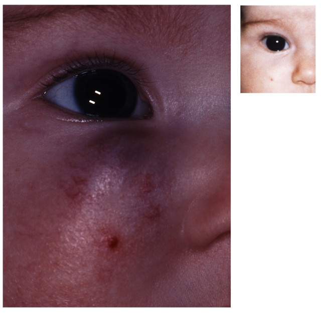
Purpose: To investigate whether neonatal dacryocystoceles and dacryocystitis are associated with nasolacrimal duct cysts, and to report the outcomes of treatment of these disorders.

Methods: This was a retrospective medical record review of two groups of infants with nasolacrimal duct (NLD) obstruction. The first group had dacryocystoceles with or without dacryocystitis. The second group had NLD obstruction with symptoms severe enough to require early NLD probing. All of the patients underwent NLD probing and nasal endoscopy. When present, NLD cysts were removed.

Results: In the first group, 33 infants had dacryocystoceles. Acute dacryocystitis was present in 16 patients, 12 had noninfected dacryocystoceles that did not resolve, and 5 had dacryocystoceles that resolved but severe symptoms persisted. All of the patients had NLD cysts that were surgically removed. The symptoms resolved after surgery in 31 patients (94%). In the second group, 27 infants less than 6 months old without dacryocystoceles underwent early NLD probing and endoscopy due to severity of symptoms. Twelve (44%) of these patients had NLD cysts. The symptoms resolved in 11 (92%) of 12 patients following NLD probing and cyst removal.

Conclusions: Neonatal dacryocystoceles are almost always associated with NLD cysts. The success rate of NLD probing and endoscopic cyst removal in these patients is excellent. Nasolacrimal duct cysts also are present in many young infants with severe symptoms of NLD obstruction. Nasal endoscopy is an important adjunct to the management of these infants.


Figures

FIGURE 1
FIGURE 1
Infant with acute dacryocystitis and dacryocystocele. Note reflux of purulent material due to application of pressure over lacrimal sac abscess.
FIGURE 2
FIGURE 2
Histological sections of nasolacrimal duct cysts. Left, Cyst lined by nasal mucosal-type pseudostratified columnar ciliated epithelium (hematoxylin and eosin, ×400). Some chronic inflammatory changes are present within the stroma. Right, Similar findings in different patient with cyst (hematoxylin and eosin, ×600).
FIGURE 3
FIGURE 3
Patient with history of nasolacrimal duct obstruction with marked erythema of upper and lower eyelids, with lesser involvement of periocular skin. The patient did not have a history of a dacryocystocele.
FIGURE 4
FIGURE 4
Case 1. Magnetic resonance image showing bilateral enlargement of the lacrimal sacs and lacrimal ducts, and nasolacrimal duct cysts (arrows), right greater than left.
FIGURE 5
FIGURE 5
Section from cadaver of infant showing cystic protrusion of distal nasolacrimal duct extending beneath the inferior turbinate (arrow on left cyst), following injection of fluid into lacrimal system. (Reprinted with permission. Archives of Ophthalmology, Volume 47, Page 156. Copyright © 1952 American Medical Association. All rights reserved.)
FIGURE 6
FIGURE 6
In order for a nasolacrimal duct cyst to form, fluid must be able to enter, but not exit, the lacrimal sac. This presumed one-way valve effect creates pressure within the lacrimal system (arrows), causing distention of the lacrimal sac and formation of a nasolacrimal duct cyst due to protrusion of the imperforate membrane occluding the distal lacrimal duct.
FIGURE 7
FIGURE 7
Dacryocystocele in an infant. Note violet-hued mass overlying left lacrimal sac, beneath the medial canthus.
FIGURE 8
FIGURE 8
Infant with infected dacryocystocele, with erythema and distention of tissue overlying the lacrimal sac, cellulitis extending onto upper eyelid, and purulent discharge.
FIGURE 9
FIGURE 9
Hemangioma overlying right lacrimal sac in a 7-month-old infant. The lesion has a vascular appearance on the surface.
FIGURE 10
FIGURE 10
Left, Four-month-old female with right periocular hemangioma. Note the vascular markings overlying the lesion and location not directly over the lacrimal sac. Right, Family photograph of patient at age 2 months shows no abnormality.
FIGURE 11
FIGURE 11
Case 3. Seventeen-month-old infant with bilateral swelling and erythema in the area of the lacrimal sacs due to ruptured dermoid cyst.
FIGURE 12
FIGURE 12
Case 4. Left, Four-month-old infant with periocular firm whitish nodule and symptoms of nasolacrimal obstruction. Right, Same patient at age 11 months with marked swelling and erythema overlying same area as previous nodule.
FIGURE 13
FIGURE 13
Case 4. Computed tomography image showing distention of lacrimal sac (arrow) extending adjacent to lower eyelid.
FIGURE 14
FIGURE 14
Left, Endoscopic view of right nasolacrimal duct cyst (arrow) after infracture of inferior nasal turbinate. Middle, The cyst is penetrated by a Bowman probe. If the cyst is not removed, a large flaccid mass remains at the distal nasolacrimal duct, which may reapproximate to cause recurrent obstruction. Right, After the probe is withdrawn, purulent material escapes from the cyst (arrow), due to presence of abscess within the lacrimal system.
FIGURE 15
FIGURE 15
Left, Initial endoscopic view of patient with right nasolacrimal duct cyst. The inferior turbinate obstructs the view of the cyst. Right, After infracture of the inferior turbinate, the nasolacrimal duct cyst is easily visualized (arrow).

References

    1. Guerry D, III, Kendig EL., Jr Congenital impatency of the nasolacrimal duct. Arch Ophthalmol. 1948;39(2):193–201. - PubMed
    1. MacEwen CJ, Young JDH. Epiphora during the first year of life. Eye (Lond) 1991;5(Pt 5):596–600. - PubMed
    1. MacEwen CJ, Phillips MG, Young JDH. Value of bacterial culturing in the course of congenital nasolacrimal duct (NLD) obstruction. J Pediatr Ophthalmol Strabismus. 1994;31(4):246–250. - PubMed
    1. Harris GJ, DiClementi D. Congenital dacryocystocele. Arch Ophthalmol. 1982;100(11):1763–1765. - PubMed
    1. Sullivan TJ, Clarke MP, Morin JD, Pashby RC. Management of congenital dacryocystocoele. Aust N Z J Ophthalmol. 1992;20(2):105–108. - PubMed