

The perception of dynamic and static facial expressions of happiness and disgust investigated by ERPs and fMRI constrained source analysis
- PMID: 23818974
- PMCID: PMC3688578
- DOI: 10.1371/journal.pone.0066997
The perception of dynamic and static facial expressions of happiness and disgust investigated by ERPs and fMRI constrained source analysis
Abstract
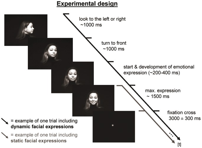
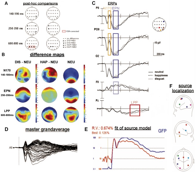
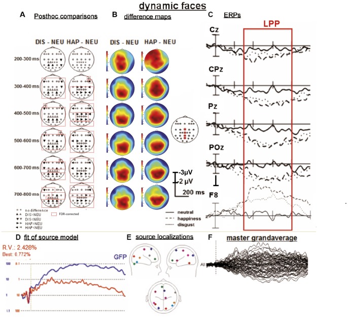
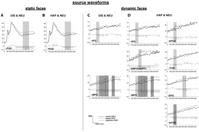
A recent functional magnetic resonance imaging (fMRI) study by our group demonstrated that dynamic emotional faces are more accurately recognized and evoked more widespread patterns of hemodynamic brain responses than static emotional faces. Based on this experimental design, the present study aimed at investigating the spatio-temporal processing of static and dynamic emotional facial expressions in 19 healthy women by means of multi-channel electroencephalography (EEG), event-related potentials (ERP) and fMRI-constrained regional source analyses. ERP analysis showed an increased amplitude of the LPP (late posterior positivity) over centro-parietal regions for static facial expressions of disgust compared to neutral faces. In addition, the LPP was more widespread and temporally prolonged for dynamic compared to static faces of disgust and happiness. fMRI constrained source analysis on static emotional face stimuli indicated the spatio-temporal modulation of predominantly posterior regional brain activation related to the visual processing stream for both emotional valences when compared to the neutral condition in the fusiform gyrus. The spatio-temporal processing of dynamic stimuli yielded enhanced source activity for emotional compared to neutral conditions in temporal (e.g., fusiform gyrus), and frontal regions (e.g., ventromedial prefrontal cortex, medial and inferior frontal cortex) in early and again in later time windows. The present data support the view that dynamic facial displays trigger more information reflected in complex neural networks, in particular because of their changing features potentially triggering sustained activation related to a continuing evaluation of those faces. A combined fMRI and EEG approach thus provides an advanced insight to the spatio-temporal characteristics of emotional face processing, by also revealing additional neural generators, not identifiable by the only use of an fMRI approach.
Conflict of interest statement
Figures
Similar articles
-
Emotions in motion: dynamic compared to static facial expressions of disgust and happiness reveal more widespread emotion-specific activations.Brain Res. 2009 Aug 11;1284:100-15. doi: 10.1016/j.brainres.2009.05.075. Epub 2009 Jun 6. Brain Res. 2009. PMID: 19501062
-
Early brain responses to affective faces: A simultaneous EEG-fMRI study.Neuroimage. 2018 Sep;178:660-667. doi: 10.1016/j.neuroimage.2018.05.081. Epub 2018 Jun 1. Neuroimage. 2018. PMID: 29864521
-
Fusiform gyrus responses to neutral and emotional faces in children with autism spectrum disorders: a high density ERP study.Behav Brain Res. 2013 Aug 15;251:155-62. doi: 10.1016/j.bbr.2012.10.040. Epub 2012 Nov 1. Behav Brain Res. 2013. PMID: 23124137
-
Distributed and interactive brain mechanisms during emotion face perception: evidence from functional neuroimaging.Neuropsychologia. 2007 Jan 7;45(1):174-94. doi: 10.1016/j.neuropsychologia.2006.06.003. Epub 2006 Jul 18. Neuropsychologia. 2007. PMID: 16854439 Review.
-
Functional atlas of emotional faces processing: a voxel-based meta-analysis of 105 functional magnetic resonance imaging studies.J Psychiatry Neurosci. 2009 Nov;34(6):418-32. J Psychiatry Neurosci. 2009. PMID: 19949718 Free PMC article. Review.
Cited by
-
Temporal Dynamics of Natural Static Emotional Facial Expressions Decoding: A Study Using Event- and Eye Fixation-Related Potentials.Front Psychol. 2018 Jul 12;9:1190. doi: 10.3389/fpsyg.2018.01190. eCollection 2018. Front Psychol. 2018. PMID: 30050487 Free PMC article.
-
Background Odors Modulate N170 ERP Component and Perception of Emotional Facial Stimuli.Front Psychol. 2018 Jun 26;9:1000. doi: 10.3389/fpsyg.2018.01000. eCollection 2018. Front Psychol. 2018. PMID: 29997539 Free PMC article.
-
Brain Responses to Emotional Faces in Natural Settings: A Wireless Mobile EEG Recording Study.Front Psychol. 2018 Oct 25;9:2003. doi: 10.3389/fpsyg.2018.02003. eCollection 2018. Front Psychol. 2018. PMID: 30410458 Free PMC article.
-
Brain synchronization during perception of facial emotional expressions with natural and unnatural dynamics.PLoS One. 2017 Jul 19;12(7):e0181225. doi: 10.1371/journal.pone.0181225. eCollection 2017. PLoS One. 2017. PMID: 28723957 Free PMC article.
-
Spatiotemporal neural network dynamics for the processing of dynamic facial expressions.Sci Rep. 2015 Jul 24;5:12432. doi: 10.1038/srep12432. Sci Rep. 2015. PMID: 26206708 Free PMC article. Clinical Trial.
References
-
- Adolphs R (2002) Recognizing emotion from facial expressions: psychological and neurological mechanisms. Behav Cogn Neurosci Rev 1: 21–62. - PubMed
-
- Posamentier MT, Abdi H (2003) Processing faces and facial expressions. Neuropsychol Rev 13: 113–143. - PubMed
-
- Vuilleumier P, Pourtois G (2007) Distributed and interactive brain mechanisms during emotion face perception: evidence from functional neuroimaging. Neuropsychologia 45: 174–194 doi:10.1016/j.neuropsychologia.2006.06.003 - DOI - PubMed
-
- Hasselmo ME, Rolls ET, Baylis GC (1989) The role of expression and identity in the face-selective responses of neurons in the temporal visual cortex of the monkey. Behav Brain Res 32: 203–218. - PubMed
-
- Allison T, Puce A, Spencer DD, McCarthy G (1999) Electrophysiological studies of human face perception. I: Potentials generated in occipitotemporal cortex by face and non-face stimuli. Cereb Cortex 9: 415–430. - PubMed
Publication types
MeSH terms
LinkOut - more resources
Full Text Sources
Other Literature Sources
Medical
