. 2013 Apr;5(2):19-39.

Blood Clotting Factor VIII: From Evolution to Therapy

Affiliations

Blood Clotting Factor VIII: From Evolution to Therapy

N A Orlova et al. Acta Naturae. 2013 Apr.

Abstract

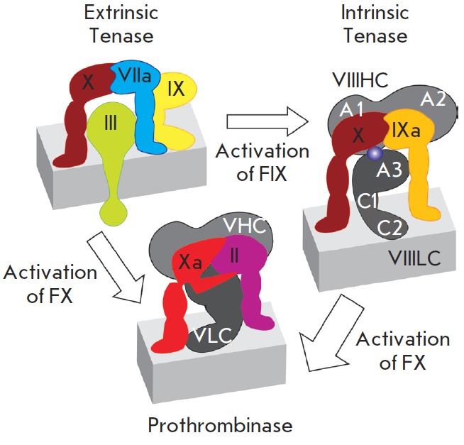
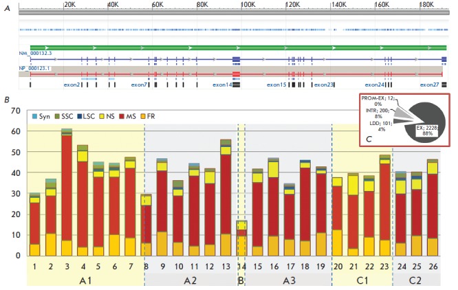
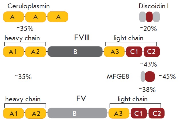
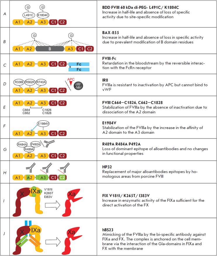
Recombinant blood clotting factor VIII is one of the most complex proteins for industrial manufacturing due to the low efficiency of its gene transcription, massive intracellular loss of its proprotein during post-translational processing, and the instability of the secreted protein. Improvement in hemophilia A therapy requires a steady increase in the production of factor VIII drugs despite tightening standards of product quality and viral safety. More efficient systems for heterologous expression of factor VIII can be created on the basis of the discovered properties of its gene transcription, post-translational processing, and behavior in the bloodstream. The present review describes the deletion variants of factor VIII protein with increased secretion efficiency and the prospects for the pharmaceutical development of longer acting variants and derivatives of factor VIII.

Keywords: blood clotting factor VIII; hemophilia A; heterologous protein expression systems.


Figures

Fig. 1
Fig. 1
Tenase complex assembly on the cell membrane. Intact proenzymes of coagulation factors are denoted by Roman numerals, activated enzymatic factors are denoted by the letter “a”. Blood clotting factors are bound to the membrane surface, Factor III is the integral membrane protein. VIIIHC – heavy chain of Factor VIII, VIIILC – light chain of Factor VIII, domains A1, A2, A3, C1, C2 of Factor VIII are denoted by white letters. VHC – heavy chain of Factor V, VLC – light chain of Factor V. The thickness of the reaction arrows corresponds to the reaction rates
Fig. 2
Fig. 2
FVIII gene structure and frequencies of the mutations causing hemophilia A. Panel A: FVIII gene on the X chromosome, NCBI reference sequence number: NG_011403.1. Transcribed are two products of alternative splicing. Functional protein FVIII is coded by the transcription variant 1, reference number of mRNA NM_000132.3, reference number of the protein NP_000123.1. Panel B: Variants of mutations in FVIII gene exons according to [3]. The number of different recorded mutations per 100 bp of the coding sequence is shown. Abbreviations: NS – nonsense mutation; MS – missense; FR – frameshift; SSC – small structural change (in-frame, < 50 bp=""); LSC="" –="" large="" structural="" change="" (>50 bp). Numbers of histogram columns correspond to the exon numbers, names of protein domains are stated below the exon numbers. Length of exon 1 in mRNA is 314 b; the only coding part of this exon (including the signal peptide), 143 b, was included in the calculations; length of exon 26 in mRNA is 1965 b, the only coding part of this exon – 156 b – was used in the calculations. The lengths of the other exons are as follows: 2 – 122 b, 3 – 123 b, 4 – 213 b, 5 – 69 b, 6 – 117 b, 7 – 222 b, 8 – 262 b, 9 – 172 b, 10 – 94 b, 11 –215 b, 12 – 151 b, 13 – 210 b, 14 – 3106 b, 15 – 154 b, 16 – 213 b, 17 – 229 b, 18 – 183 b, 19 – 117 b, 20 – 72 b, 21 – 86 b, 22 – 156 b, 23 – 145 b, 24 – 149 b, 25 – 177 b. Panel C: Variants of mutations in the FVIII gene. Abbreviations: LDD – large deletions and duplications in one or multiple domains of FVIII; INTR – distortions in the splice sites; PROM-EX – promoter area mutations and deletions in the promoter area plus the exon; EX – mutations in the exons. Primary data taken from [3], extracted July 18, 2012
Fig. 3
Fig. 3
Domain structure of FVIII homologues. Numbers represent the homology level of amino acids for domain groups. Discoidin I was obtained from D. discoideum; all other proteins were obtained from H. sapiens
Fig. 4
Fig. 4
3D structure of the FVIII deletion variant according to [42]. Core residues of N-linked glycans are shown in red, FVIII heavy chain is shown in blue, light chain is shown in green
Fig. 5
Fig. 5
Post-translational modifications and functional sites of FVIII. N-glycosylation sites are denoted by circles. Filled black circles – occupied sites, half-filled circle – partially occupied site, filled gray circle – presumably occupied site, red circles – unoccupied sites. Disulphide bonds are denoted by brackets, gray bracket – presumably existing disulphide bond. Red vertical lines – reduced Cys residues, the actual state of Cys residues in the B domain is unknown. S inside a circle – sulfated Tyr residues. Light blue marks the areas of interaction with corresponding clotting factors, phospholipids (Pl), von Willebrand factor (vWF), and copper ions (Cu+). SP – signal peptide and propeptide
Fig. 6
Fig. 6
Intracellular traffic of the FVIII polypeptide to be secreted. OST – Oligosaccharyltransferase, Sec61 – membrane protein translocator, GI and GII – glucosidases I and II; CNX – calnexin, CRT – calreticulin, GRP78/BiP – glucose regulated protein 78 / immunoglobulin binding protein; GRP94 – glucose regulated protein 94; PDI, ERp57 – disulphide isomerases; MCFD2 – multiple coagulation factors deficiency protein 2; LMAN1 – mannose-binding lectin 1; COPI, COPII – vesicle coat proteins I and II; ERGIC – ER-Golgi intermediate compartment
Fig. 7
Fig. 7
Proteolytic processing of FVIII. Black triangles – sites of processing by the PACE/furin family proteases; green triangles – sites of processing during activation; red triangles – sites of processing during inactivation; and the blue circle represents the coordinated copper ion(s)
Fig. 8
Fig. 8
FVIII long-acting variants and functional mimetics. Spirals represent the covalently attached PEG groups; dashed lines – unknown conjugation sites; arrows – noncovalent interaction; and wavy lines – blocking of interactions. Protein parts from porcine FVIII are shown in green in panel H

References

    1. Antonarakis S.E., Kazazian H.H., Tuddenham E.G.. Hum. Mutat. 1995;5(1):1–22. - PubMed
    1. A Mutation Database. http://hadb.org.uk. Kemball-Cook G., Haemophilia A., University College London. 1994
    1. Payne A.B., Miller C.H., Kelly F.M., Michael Soucie J., Craig Hooper W., Hum. Mutat. 2013;34(2):2382–2391. - PMC - PubMed
    1. Blumel J., Schmidt I., Effenberger W., Seitz H., Willkommen H., Brackmann H.H., Lower J., Eis-Hubinger A.M.. Transfusion. 2002;42(11):1473–1481. - PubMed
    1. Yokozaki S., Fukuda Y., Nakano I., Katano Y., Toyoda H., Takamatsu J.. Blood. 1999;94(10):3617. - PubMed

LinkOut - more resources