

Acute gastric dilatation: a transient cause of hepatic portal venous gas-case report and review of the literature
- PMID: 23819076
- PMCID: PMC3683454
- DOI: 10.1155/2013/723160
Acute gastric dilatation: a transient cause of hepatic portal venous gas-case report and review of the literature
Abstract
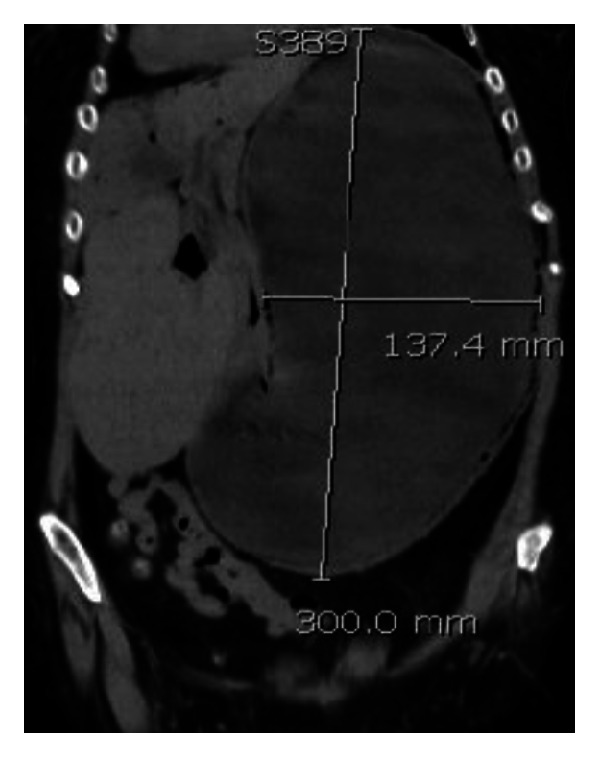
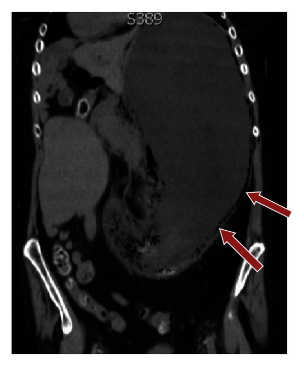
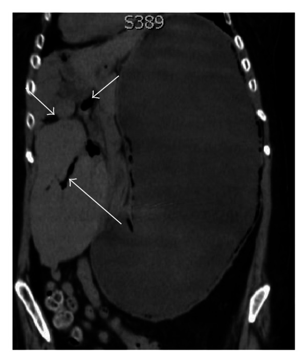
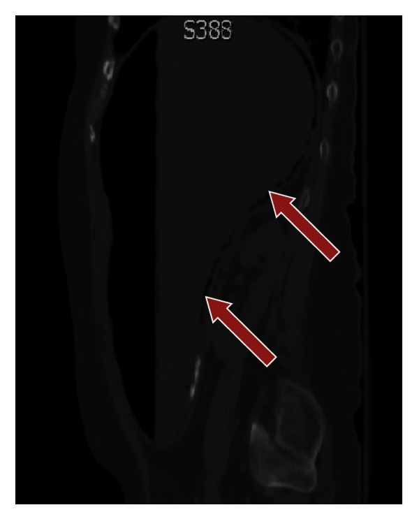
Gastric pneumatosis (GP) and hepatic portal venous gas (HPVG) have typically been thought of as an ominous radiological sign associated with a grave prognosis, and the observation of HPVG on plain abdominal radiography, ultrasonography, or computed tomography is viewed as a significant finding. It is often associated with severe or potentially lethal conditions warranting urgent diagnosis and possible surgical intervention. Early studies of HPVG based on plain abdominal radiography found an associated mortality rate of 75% primarily due to ischemic bowel. However, modern abdominal computed tomography (CT) has resulted in the detection of HPVG in an increased proportion of nonfatal and benign conditions. We report a nonfatal case of HPVG in a patient with Noonan's syndrome due to acute gastric dilatation in the setting of gastric outlet obstruction caused by a congenital band that is extremely rare in adults.
Figures
References
-
- Wolfe JN, Evans WA. Gas in the portal veins of the liver in infants; a roentgenographic demonstration with postmortem anatomical correlation. The American Journal of Roentgenology, Radium Therapy, and Nuclear Medicine. 1955;74(3):486–488. - PubMed
-
- Susman N, Senturia HR. Gas embolization of the portal venous system. The American Journal of Roentgenology, Radium Therapy, and Nuclear Medicine. 1960;83:847–850. - PubMed
-
- Benson MD. Adult survival with intrahepatic portal venous gas secondary to acute gastric dilatation, with a review of portal venous gas. Clinical Radiology. 1985;36(4):441–443. - PubMed
-
- Faberman RS, Mayo-Smith WW. Outcome of 17 patients with portal venous gas detected by CT. American Journal of Roentgenology. 1997;169(6):1535–1538. - PubMed
-
- Iannitti DA, Gregg SC, Mayo-Smith WW, Tomolonis RJ, Cioffi WG, Pricolo VE. Portal venous gas detected by computed tomography: is surgery imperative? Digestive Surgery. 2003;20(4):306–315. - PubMed
LinkOut - more resources
Full Text Sources
Other Literature Sources
