. 2013 Nov;15(11):2937-50.
doi: 10.1111/1462-2920.12179. Epub 2013 Jul 3.

The first engagement of partners in the Euprymna scolopes-Vibrio fischeri symbiosis is a two-step process initiated by a few environmental symbiont cells

Affiliations

The first engagement of partners in the Euprymna scolopes-Vibrio fischeri symbiosis is a two-step process initiated by a few environmental symbiont cells

Melissa A Altura et al. Environ Microbiol. 2013 Nov.

Abstract

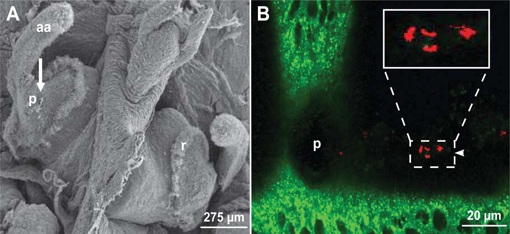
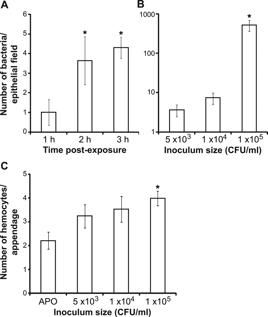
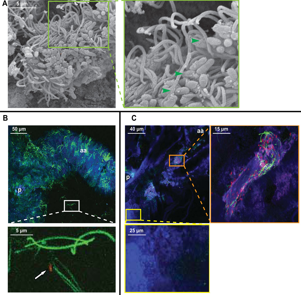
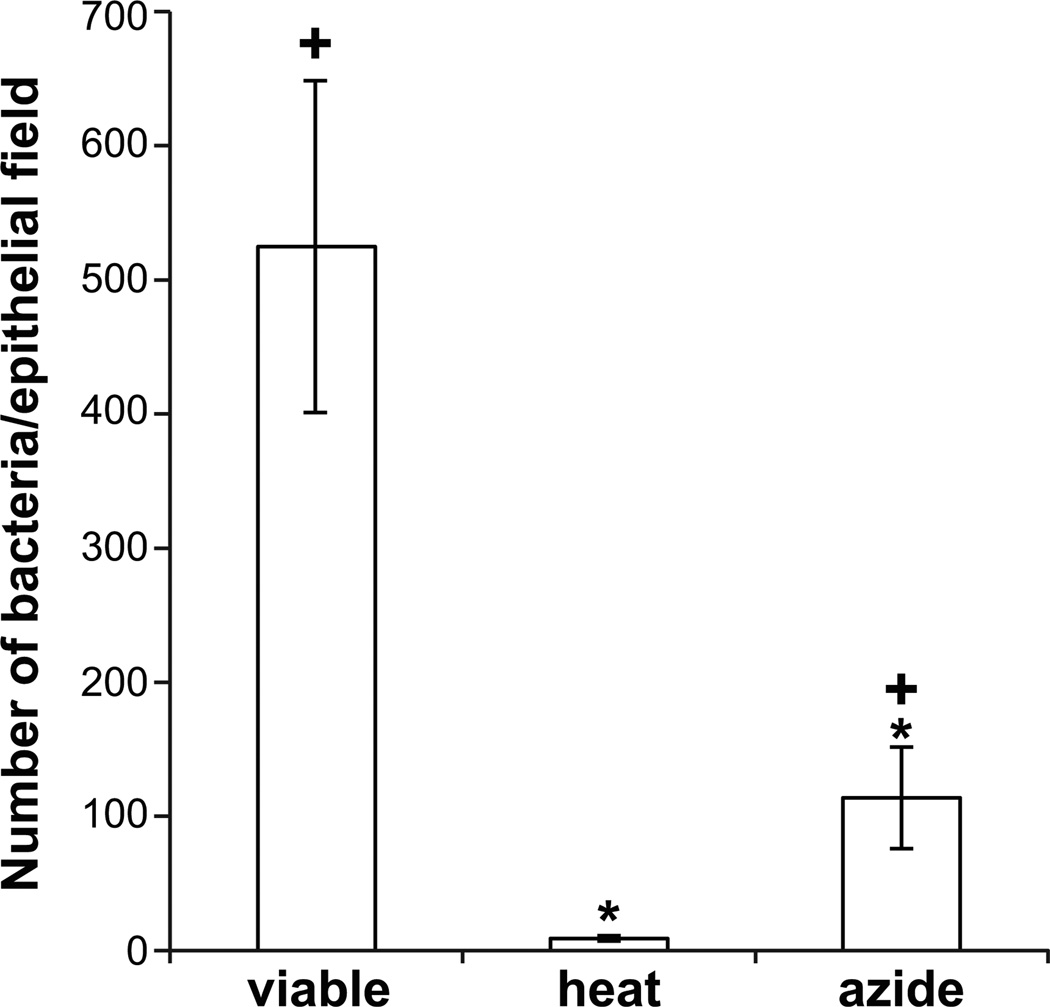
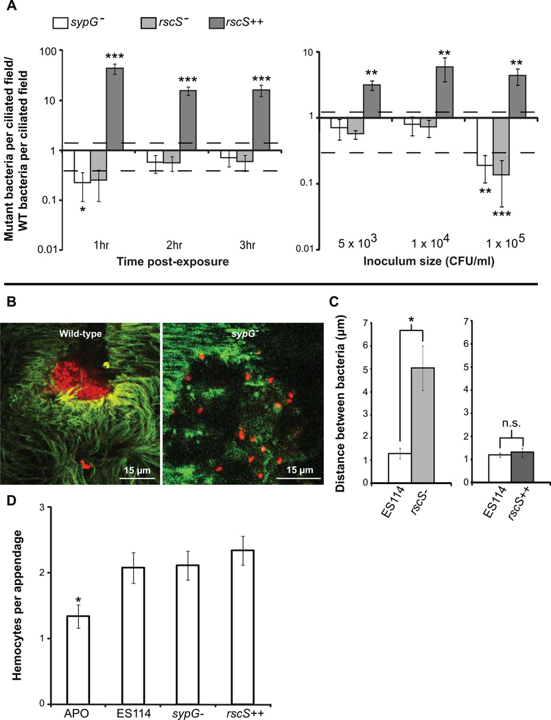
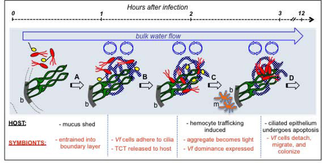
We studied the Euprymna scolopes-Vibrio fischeri symbiosis to characterize, in vivo and in real time, the transition between the bacterial partner's free-living and symbiotic life styles. Previous studies using high inocula demonstrated that environmental V. fischeri cells aggregate during a 3 h period in host-shed mucus along the light organ's superficial ciliated epithelia. Under lower inoculum conditions, similar to the levels of symbiont cells in the environment, this interaction induces haemocyte trafficking into these tissues. Here, in experiments simulating natural conditions, microscopy revealed that at 3 h following first exposure, only ∼ 5 V. fischeri cells aggregated on the organ surface. These cells associated with host cilia and induced haemocyte trafficking. Symbiont viability was essential and mutants defective in symbiosis initiation and/or production of certain surface features, including the Mam7 protein, which is implicated in host cell attachment of V. cholerae, associated normally with host cilia. Studies with exopolysaccharide mutants, which are defective in aggregation, suggest a two-step process of V. fischeri cell engagement: association with host cilia followed by aggregation, i.e. host cell-symbiont interaction with subsequent symbiont-symbiont cell interaction. Taken together, these data provide a new model of early partner engagement, a complex model of host-symbiont interaction with exquisite sensitivity.


Conflict of interest statement

The authors declare no conflict of interest.

Figures

Fig. 1
Fig. 1
The site of initial association of environmental V. fischeri with host tissues. A. An SEM of the juvenile squid light organ. Ciliated epithelial fields, which occur on the lateral faces of the organ, are false-colored in green; arrow, location in panel B of aggregating symbionts. B. A confocal image of the ciliated epithelium (cytoplasm counterstained with CellTracker Green) of a living animal. An aggregate of 5 live RFP-labeled V. fischeri (red) associated with the light organ surface (not all are in this focal plane shown). White box, higher magnification of the associating V. fischeri bacteria. aa = anterior appendage, p = pore, r = ciliated ridge.
Fig. 2
Fig. 2
Quantification of V. fischeri ES114 cells in ciliated epithelial fields of juvenile light organs. [Representative graphs; all experiments replicated at least twice] A. The effect of varying exposure time (h = hours) on the number of wild-type V. fischeri per ciliated field after exposure to an environmentally-relevant dose of 5 × 10³ CFU/ml. *, data points that were significantly different from 1 h post-exposure, but not from one another (Mann-Whitney test with a Bonferroni correction for multiple comparisons; n = 5 independent sample animals for all conditions). B. The effect of varying inoculum size on the number of V. fischeri cells per ciliated field at a constant exposure time of 3 h. *, data points that were significantly different from 5 × 10³ CFU/ml (Mann-Whitney test with a Bonferroni correction for multiple comparisons; n = 5 independent sample animals for all conditions). C. The effect of varying inoculum size on the average number of hemocytes trafficked to the blood sinus space underlying the ciliated epithelium of the light organ. *, data points that were significantly different from the aposymbiotic (Poisson p-value analysis; APO, n = 9; 5 × 10³, n = 8; 1 × 10⁴, n=11; 1 × 10⁵, n = 11). Bars, standard error.
Fig. 3
Fig. 3
Representative micrographs of V. fischeri associated with the ciliated epithelium of the juvenile light organ. A. An SEM micrograph of V. fischeri cells (false colored) associating with the organ surface of an animal exposed to 1×10⁶ symbiont CFU/ml and then fixed. Green arrows indicate examples of bacterial-ciliary contacts. B. Upper: confocal micrograph of a live animal specimen exposed to 5 × 10³ CFU/ml V. fischeri (red) for 3 h. Box, area magnified in lower panel. Lower: RFP-expressing V. fischeri associating with a host cilium. [Counterstains: cilia, TubulinTracker (green); mucus, wheat germ agglutinin (blue); image is highly pixelated due to use of living tissue, which requires acquiring images at high scan speeds] C. Upper, left: confocal micrograph of a live animal specimen exposed to 1 × 10⁵ CFU/ml V. fischeri (red) for 3 h. Orange box, area magnified in upper right. Yellow box, area magnified in lower panel. Upper, right: higher magnification of RFP-expressing V. fischeri associating with host cilia. Lower: area of mucus without cilia, which contains no V. fischeri. [Counterstains: cilia, TubulinTracker (green); mucus, wheat germ agglutinin (blue)]
Fig 4
Fig 4
The effect of V. fischeri viability on association with the ciliated epithelial field of the juvenile light organ. [Representative graph; all experiments replicated at least twice] Number of bacteria/ciliated field after 3 h exposure to 1 × 10⁵ CFU/ml of viable, heat-killed, or azide-killed V. fischeri. *, data points that were significantly different from viable bacteria and +, data points that were significantly different from heat-killed bacteria, both according to a Mann-Whitney test with a Bonferroni correction for multiple comparisons (n=5 independent sample animals for all conditions). Bars, standard error.
Fig 5
Fig 5
The two-step process of the host’s engagement of V. fischeri cells. [Representative graphs; all experiments replicated at least twice] A. Left: The effect of varying time on the number of mutant V. fischeri bacteria/ciliated field after exposure to an environmentally relevant dose of 5 × 10³ CFU/ml. Both strain and time post-exposure to bacteria affected variation in the RscS++ colonization data as tested by a 2-way ANOVA, time, p<0.05, and strain, p<0.0001. Right: The effect of varying inoculum size on the number of mutant V. fischeri bacteria/ciliated field at a constant exposure time of 3 h (n=5 independent sample animals for all conditions). For SypG- and RscS-strain experiments, all three factors (Strain, Inoculum, and the interaction factor) were significant with p < 0.05 by a 2-way ANOVA. For RscS++ strain experiments only strain accounted for variation in the data by a 2-way ANOVA, p<0.0001. Dashed line, standard error for wild-type controls. Significant differences from wild-type are denoted as follows: *, p-value < 0.05, **, p-value < 0.01, ***, p-value < 0.001 by a Dunn’s post-hoc pairwise comparison within each strain’s data set after a Bonferroni post-test for multiple comparisons. B. Confocal micrographs of RFP-expressing V. fischeri wild type or sypG bacteria in association with the ciliated epithelium [Counterstain: cilia, TubulinTracker (green)] C. The effect of altered exopolysaccharide production on the average distance between aggregating bacteria. (n=10 pairs of bacteria from 3 independent sample animals for all conditions; inoculation, 1×10⁵ CFU/ml seawater). *, p-value < 0.05, data points that were significantly different from WT/ES114 according to a Mann-Whitney test. Bars, standard error. D. The amount of hemocyte trafficking to the blood sinus space of the light organ anterior appendage due to exposure to exopolysaccharide mutants. (APO, n=44; ES114, n=38; sypG, n=44; rscS++, n=47; inoculation 104 CFU/ml of seawater) *, indicates that the average of the two distributions is significantly different with a Poissonian p-value of <0.05. Bars, standard error.
Fig 6
Fig 6
The ability of non-symbiotic vibrios to associate with the ciliated epithelial field of the juvenile light organ. [Representative graphs; all experiments replicated at least twice] A. Left: The effect of different lengths of exposure on the number of V. fischeri MJ11 and V. parahaemolyticus KNH1 bacteria/ciliated field after exposure to an inoculum of 5 × 10³ CFU/ml. Right: The effect of inoculum size on the number of V. fischeri MJ11 or V. parahaemolyticus KNH1 cells per ciliated field after a constant exposure time of 3 h. Dashed line, standard error for V. fischeri ES114 controls. Data were analyzed by a 2-way ANOVA followed by a Bonferroni post-test for multiple comparisons, though no significant differences were found. B. The effect of increasing V. fischeri ES114 inoculum size on the number of V. parahaemolyticus KNH1 bacteria/ciliated field. Exposure time, 3 h. KNH1 inoculum, 5 × 10³ CFU/ml. C. The effect of V. fischeri aggregation on the ability of V. parahaemolyticus KNH1 to compete with V. fischeri. Exposure time, 2 h. (n=5 independent sample animals for all conditions). Bars, standard error.
Fig. 7
Fig. 7
A new model of the initial events of partner interaction in the squid-vibrio association. The early events can be defined as a series of transitions, A–D. Immediately upon hatching, water harboring environmental bacteria (red, V. fischeri; yellow, non-specific Gram-negative) is brought through the host’s body cavity into the vicinity of the cilia (green) on the surface (thick grey line where cilia attach) of the nascent symbiotic organ. Mucus (blue mottling) is shed (transition A), Gram-negative bacteria then bind to the cilia (transition B), V. fischeri releases TCT and host responds with the trafficking of macrophage-like blood cells (m) into the blood sinus (b) under the ciliated epithelium (transition C), and finally V. fischeri migrates into host tissues and colonizes deep crypt spaces of the organ, where it induces host morphogenesis (transition D). [See text for details.]

References

    1. Adam EC, Mitchell BS, Schumacher DU, Grant G, Schumacher U. Pseudomonas aeruginosa II lectin stops human ciliary beating: therapeutic implications of fucose. Am J Respir Crit Care Med. 1997;155:2102–2104. - PubMed
    1. Aeckersberg F, Lupp C, Feliciano B, Ruby EG. Vibrio fischeri outer membrane protein OmpU plays a role in normal symbiotic colonization. J Bacteriol. 2001;183:6590–6597. - PMC - PubMed
    1. Altura MA, Stabb E, Goldman W, Apicella M, McFall-Ngai MJ. Attenuation of host NO production by MAMPs potentiates development of the host in the squid-vibrio symbiosis. Cell Microbiol. 2011;13:527–537. - PMC - PubMed
    1. Anderton TL, Maskell DJ, Preston A. Ciliostasis is a key early event during colonization of canine tracheal tissue by Bordetella bronchiseptica. Microbiology. 2004;150:2843–2855. - PubMed
    1. Bajolet-Laudinat O, Girod-de Bentzmann S, Tournier JM, Madoulet C, Plotkowski MC, Chippaux C, Puchelle E. Cytotoxicity of Pseudomonas aeruginosa internal lectin PA-I to respiratory epithelial cells in primary culture. Infect Immun. 1994;62:4481–4487. - PMC - PubMed

Publication types

Substances

LinkOut - more resources