

The importance of a single primary cilium
- PMID: 23819944
- PMCID: PMC3812286
- DOI: 10.4161/org.25144
The importance of a single primary cilium
Abstract
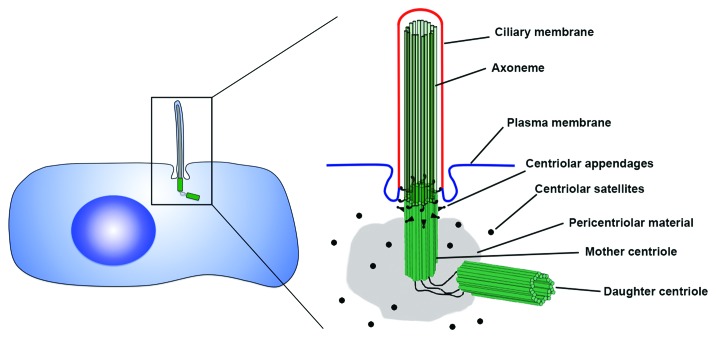
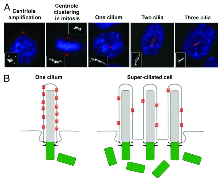
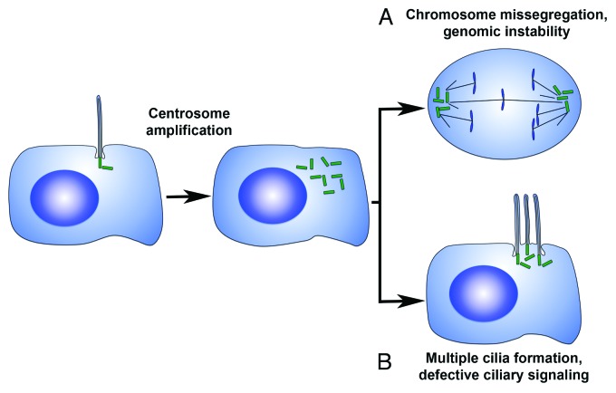
The centrosome is the main microtubule-organizing center in animal cells, and helps to influence the morphology of the microtubule cytoskeleton in interphase and mitosis. The centrosome also templates the assembly of the primary cilium, and together they serve as a nexus of cell signaling that provide cells with diverse organization, motility, and sensory functions. The majority of cells in the human body contain a solitary centrosome and cilium, and cells have evolved regulatory mechanisms to precisely control the numbers of these essential organelles. Defects in the structure and function of cilia lead to a variety of complex disease phenotypes termed ciliopathies, while dysregulation of centrosome number has long been proposed to induce genome instability and tumor formation. Here, we review recent findings that link centrosome amplification to changes in cilium number and signaling capacity, and discuss how supernumerary centrosomes may be an important aspect of a set of cilia-related disease phenotypes.
Keywords: cancer; centriole; centrosome; ciliopathy; cilium.
Figures
Similar articles
-
The Centrosome and the Primary Cilium: The Yin and Yang of a Hybrid Organelle.Cells. 2019 Jul 10;8(7):701. doi: 10.3390/cells8070701. Cells. 2019. PMID: 31295970 Free PMC article. Review.
-
Mechanism and Regulation of Centriole and Cilium Biogenesis.Annu Rev Biochem. 2019 Jun 20;88:691-724. doi: 10.1146/annurev-biochem-013118-111153. Epub 2019 Jan 11. Annu Rev Biochem. 2019. PMID: 30601682 Free PMC article. Review.
-
Oncogenic FGFR Fusions Produce Centrosome and Cilia Defects by Ectopic Signaling.Cells. 2021 Jun 9;10(6):1445. doi: 10.3390/cells10061445. Cells. 2021. PMID: 34207779 Free PMC article. Review.
-
Primary Cilia Reconsidered in the Context of Ciliopathies: Extraciliary and Ciliary Functions of Cilia Proteins Converge on a Polarity theme?Bioessays. 2018 Aug;40(8):e1700132. doi: 10.1002/bies.201700132. Epub 2018 Jun 8. Bioessays. 2018. PMID: 29882973 Free PMC article. Review.
-
A Dynamic Protein Interaction Landscape of the Human Centrosome-Cilium Interface.Cell. 2015 Dec 3;163(6):1484-99. doi: 10.1016/j.cell.2015.10.065. Cell. 2015. PMID: 26638075 Free PMC article.
Cited by
-
Polo-like kinase 4 maintains centriolar satellite integrity by phosphorylation of centrosomal protein 131 (CEP131).J Biol Chem. 2019 Apr 19;294(16):6531-6549. doi: 10.1074/jbc.RA118.004867. Epub 2019 Feb 25. J Biol Chem. 2019. PMID: 30804208 Free PMC article.
-
Scanning electron microscopy of human islet cilia.Proc Natl Acad Sci U S A. 2023 May 30;120(22):e2302624120. doi: 10.1073/pnas.2302624120. Epub 2023 May 19. Proc Natl Acad Sci U S A. 2023. PMID: 37205712 Free PMC article.
-
Cooperation between primary cilia signaling and integrin receptor extracellular matrix engagement regulates progenitor proliferation and neuronal differentiation in the developing cerebellum.Front Cell Dev Biol. 2023 Feb 21;11:1127638. doi: 10.3389/fcell.2023.1127638. eCollection 2023. Front Cell Dev Biol. 2023. PMID: 36895790 Free PMC article. Review.
-
KIFCI, a novel putative prognostic biomarker for ovarian adenocarcinomas: delineating protein interaction networks and signaling circuitries.J Ovarian Res. 2014 May 12;7:53. doi: 10.1186/1757-2215-7-53. eCollection 2014. J Ovarian Res. 2014. PMID: 25028599 Free PMC article.
-
Primary cilia in rat mature Müller glia: downregulation of IFT20 expression reduces sonic hedgehog-mediated proliferation and dedifferentiation potential of Müller glia primary cultures.Cell Mol Neurobiol. 2015 May;35(4):533-42. doi: 10.1007/s10571-014-0149-3. Epub 2014 Dec 14. Cell Mol Neurobiol. 2015. PMID: 25504432 Free PMC article.
References
Publication types
MeSH terms
Grants and funding
LinkOut - more resources
Full Text Sources
Other Literature Sources