. 2013 Sep;346(3):381-92.
doi: 10.1124/jpet.113.204958. Epub 2013 Jul 2.

The novel anticancer agent JNJ-26854165 induces cell death through inhibition of cholesterol transport and degradation of ABCA1

Affiliations

The novel anticancer agent JNJ-26854165 induces cell death through inhibition of cholesterol transport and degradation of ABCA1

Richard J Jones et al. J Pharmacol Exp Ther. 2013 Sep.

Erratum in

  • J Pharmacol Exp Ther. 2013 Nov;347(2):540

Abstract

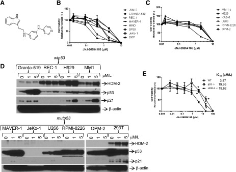
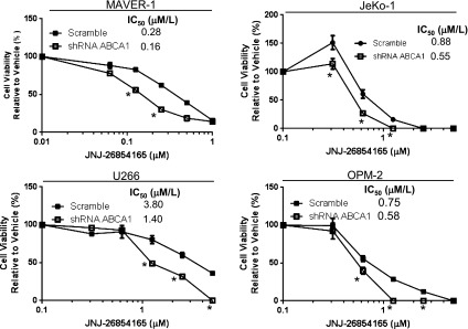
JNJ-26854165 (serdemetan) has previously been reported to inhibit the function of the E3 ligase human double minute 2, and we initially sought to characterize its activity in models of mantle cell lymphoma (MCL) and multiple myeloma (MM). Serdemetan induced a dose-dependent inhibition of proliferation in both wild-type (wt) and mutant (mut) p53 cell lines, with IC50 values from 0.25 to 3 μM/l, in association with an S phase cell cycle arrest. Caspase-3 activation was primarily seen in wtp53-bearing cells but also occurred in mutp53-bearing cells, albeit to a lesser extent. 293T cells treated with JNJ-26854165 and serdemetan-resistant fibroblasts displayed accumulation of cholesterol within endosomes, a phenotype reminiscent of that seen in the ATP-binding cassette subfamily A member-1 (ABCA1) cholesterol transport disorder, Tangiers disease. MM and MCL cells had decreased cholesterol efflux and electron microscopy demonstrated the accumulation of lipid whorls, confirming the lysosomal storage disease phenotype. JNJ-26854165 induced induction of cholesterol regulatory genes, sterol regulatory element-binding transcription factor-1 and -2, liver X receptors α and β, along with increased expression of Niemann-Pick disease type-C1 and -C2. However, JNJ-26854165 induced enhanced ABCA1 turnover despite enhancing transcription. Finally, ABCA1 depletion resulted in enhanced sensitivity to JNJ-26854165. Overall, these findings support the hypothesis that serdemetan functions in part by inhibiting cholesterol transport and that this pathway is a potential new target for the treatment of MCL and MM.


Figures

Fig. 1.
Fig. 1.
JNJ-26854165 acts independent of HDM-2 in MCL and MM cell lines. (A) Chemical structure of JNJ-26854165. MCL (B) and MM (C) cell lines were seeded in 96-well plates for viability analyses using WST-1 and treated with JNJ-26854165 for 72 hours. Results are expressed as the percentage of cell viability in relation to the vehicle-treated sample for each cell line, which was arbitrarily set at 100%. (D) Immunoblot analysis was performed on MCL and MM cells treated with serdemetan or dimethylsulfoxide for 48 hours. Protein extracts were probed for their content of p53, HDM-2, and β-actin as a loading control. A representative Western blot is shown of triplicate experiments. (E) MEFs were evaluated for sensitivity to serdemetan using WST-1 as indicated above. Experiments were done in triplicate, and the S.E.M. is shown for each point. An unpaired t test was used to determine statistical significance of p53 and HDM-2 −/− cells compared with the WT MEF control. *P values < 0.05.
Fig. 2.
Fig. 2.
JNJ-26854165 induces an S-phase arrest and cell death. MM and MCL cells with wtp53 and mutp53 were treated with IC50 concentrations (determined from the WST-1 assay in Fig. 1) of JNJ-26854165 for 48 hours, followed by cell cycle analysis along with caspase-3 and Annexin-V cell death assays. (A) Cell cycle analysis of MM and MCL cells. (B) Viable cell numbers were determined using Annexin-V/TO-PRO exclusion, cell death was determined by counting Annexin-V/ TO-PRO-positive cells and caspase-3 activity was measured using the substrate Red-DEVD-FMK. All experiments were performed in triplicate and a representative profile is shown. DMSO, dimethylsulfoxide. *P < 0.05 relative to the vehicle control.
Fig. 3.
Fig. 3.
Inhibition of cholesterol transport occurs with JNJ-26854165 treatment. (A) WT MEFs and JNJ-26854165-resistant MEFs (165R) were visualized by light microscopy (40× magnification). (B) Fluorescent staining of cholesterol using Filipin in WT and 165R MEFs (40× magnification). (C) 293T cells were treated for 24 hours with JNJ-26854165, U18666A, or pimozide, and stained with Filipin to determine cholesterol localization (40× magnification). Representative images are shown in both panels from one of three independent experiments.
Fig. 4.
Fig. 4.
Decreased cholesterol efflux and transport induced by JNJ-26854165 in MM and MCL cells. (A) MM and MCL cell lines were treated with 5 μM/l JNJ-26854165 for 24 hours stained with Filipin and cholesterol localization determined using an ImageStreamX Mark II (60× magnification). (B) MCL, MM, and 293T cell lines were treated with 5 μM/l JNJ-26854165 for 24 hours and loaded with NBD-cholesterol, and cholesterol efflux was monitored using a fluorescent plate reader for 24 hours using apolipoprotein A-I as an acceptor. DMSO, dimethylsulfoxide. *P < 0.05 relative to vehicle.
Fig. 5.
Fig. 5.
JNJ-26854165 induces lipid whorl accumulation similar to that seen in lysosomal storage disease. MM1.S and RPMI-8226 cells were treated for 48 hours with IC50 concentrations of JNJ-26854165 and then analyzed by transmission electron microscopy (7,500 and 25,000× magnification). Black arrows indicate LW, MITO indicates mitochondria, and CNT indicates centriole.
Fig. 6.
Fig. 6.
Modulation of cholesterol regulatory genes and cholesterol transporters is induced by JNJ-26854165. U266 and JeKo-1 cells were treated with JNJ-26854165 and cells harvested at the indicated time points. (A) RNA was extracted and cDNA was synthesized. The cholesterol regulatory genes, SREBF-1 and -2, and LXR-α and -β were measured, along with the cholesterol transporters NPC-1, NPC-2, and ABCA1, by quantitative real-time PCR using the ΔΔCT method, with the vehicle treated cells used as a relative calibrator and values expressed as relative transcript expression. *P < 0.05 compared with the 0 hour time point using an unpaired t test. (B) Immunoblotting of protein lysates from harvested cells at the indicated time points and probed with the indicated sera. Densitometry values for immunoblotting are shown in Supplemental Table 2. (C) JeKo-1 cells were treated with 2 μM/l JNJ-26854165 (165) or DMSO in combination with cycloheximide and harvested at the indicated time points. Protein lysates were immunoblotted for ABCA1, and expression levels were determined using densitometry. Relative ABCA1 half-life normalized to the NT (no treatment) control is shown. *P < 0.05 comparing vehicle to JNJ-26854165-treated cells using an unpaired t test. (D) 293T, WT, and 165R MEF cells were treated with 5 μM/l JNJ-26854165 for 24 hours, and lysates evaluated for the indicated cholesterol regulatory genes. All experiments were performed in triplicate and a representative profile is shown. Values represent the mean ± standard error from three independent experiments. CHX, cycloheximide; DMSO, dimethylsulfoxide; RQ, relative quantification.
Fig. 7.
Fig. 7.
Depletion of ABCA1 expression enhances sensitivity to JNJ-26854165. HeLa cells expressing a tetracycline-off regulatable ABCA1–green fluorescent protein (GFP) were maintained in the absence of doxycycline (DOXY) or with DOXY to suppress ABCA1 expression. (A) Cell viability of HeLa ABCA1-GFP cells treated with JNJ-26854165 (165) for 3 days in the presence of vehicle or DOXY (1 μg/ml). (B) HeLa ABCA1-GFP cells were treated for 3 days with JNJ-26854165 in the presence or absence of 1 μg/ml DOXY, and fluorescence microscopy images were captured at 40× magnification. (C) doxycycline (DOXY)-treated HeLa ABCA1-GFP cells were treated 24 hours with 165 stained with Filipin, cholesterol localization was determined using an ImageStreamX Mark II (60× magnification). (D) Cholesterol efflux was measured in ABCA1-GFP-depleted HeLa cells and compared with the control cells treated with 165 using NBD-cholesterol. *P < 0.05 relative to vehicle. (E) ABCA1-GFP-depleted HeLa cells treated with 165 for 24 hours and immunoblotted for ABCA1, NPC1, and NPC2. DMSO, dimethylsulfoxide; TET, tetracycline.
Fig. 8.
Fig. 8.
Suppression of ABCA1 expression enhances JNJ-26854165 sensitivity in MCL and MM cell lines. MCL and MM cells with stable shRNA knockdown of ABCA1 were treated with JNJ-26854165 for 3 days, and cell viability was determined. All experiments were performed in triplicate, and an unpaired t test was performed on indicated panels compared with the control, and P values < 0.05 are indicated with *.

References

    1. Arts J, Page M, Valckx A, Blattner C, Kulikov R, Floren W, van Nuffel L, Janssen L, King P, Masure S, et al.(2008) JNJ-26854165—a novel hdm2 antagonist in clinical development showing broad-spectrum preclinical antitumor activity against solid malignancies, in AACR Meeting Abstracts 2008 (1_Annual_Meeting): 1592.
    1. Assmann GVEA, Brewer HB (1995) Familial high density lipoprotein deficiency: Tangier disease, in The Metabolic and Molecular Bases of Inherited Disease (Scriver CR, Beaudet AL, Sly WS, and Valle D eds.) pp 2053-2072, McGraw Hill, New York.
    1. Barboza JA, Iwakuma T, Terzian T, El-Naggar AK, Lozano G. (2008) Mdm2 and Mdm4 loss regulates distinct p53 activities. Mol Cancer Res 6:947–954 - PMC - PubMed
    1. Börnig H, Geyer G. (1974) Staining of cholesterol with the fluorescent antibiotic “filipin”. Acta Histochem 50:110–115 - PubMed
    1. Boyd RS, Jukes-Jones R, Walewska R, Brown D, Dyer MJ, Cain K. (2009) Protein profiling of plasma membranes defines aberrant signaling pathways in mantle cell lymphoma. Mol Cell Proteomics 8:1501–1515 - PMC - PubMed

Publication types

MeSH terms