

Improved rat spinal cord injury model using spinal cord compression by percutaneous method
- PMID: 23820159
- PMCID: PMC3788159
- DOI: 10.4142/jvs.2013.14.3.329
Improved rat spinal cord injury model using spinal cord compression by percutaneous method
Abstract
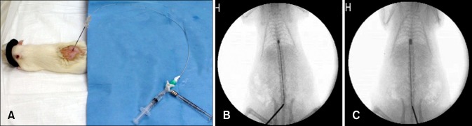
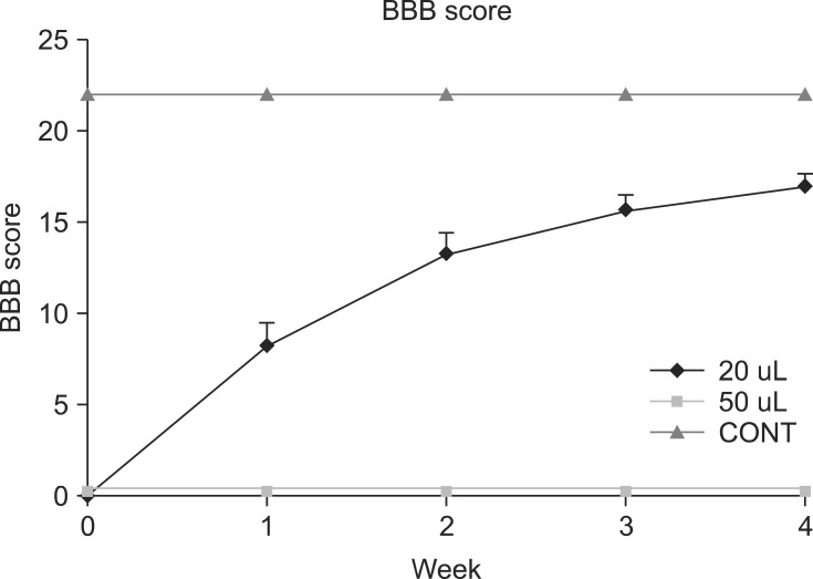
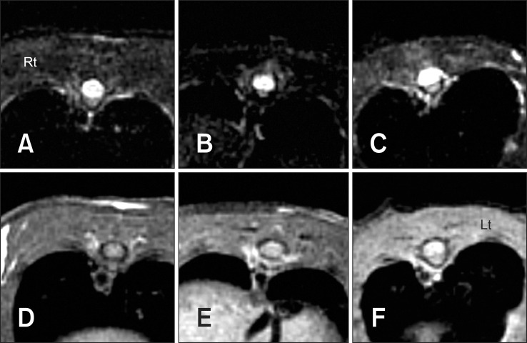
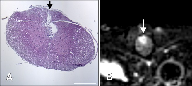
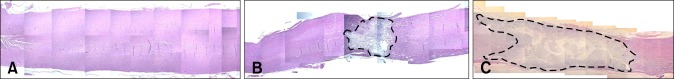
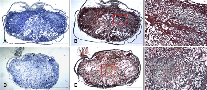
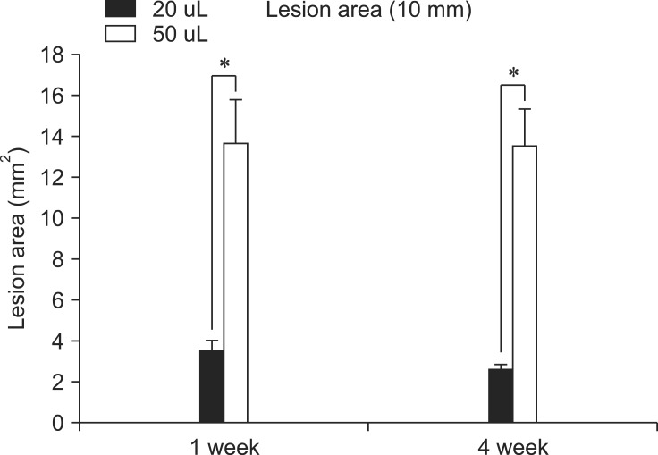
Here, percutaneous spinal cord injury (SCI) methods using a balloon catheter in adult rats are described. A balloon catheter was inserted into the epidural space through the lumbosacral junction and then inflated between T9-T10 for 10 min under fluoroscopic guidance. Animals were divided into three groups with respect to inflation volume: 20 μL (n = 18), 50 μL (n = 18) and control (Fogarty catheter inserted but not inflated; n = 10). Neurological assessments were then made based on BBB score, magnetic resonance imaging and histopathology. Both inflation volumes produced complete paralysis. Gradual recovery of motor function occurred when 20 μL was used, but not after 50 μL was applied. In the 50 μL group, all gray and white matter was lost from the center of the lesion. In addition, supramaximal damage was noted, which likely prevented spontaneous recovery. This percutaneous spinal cord compression injury model is simple, rapid with high reproducibility and the potential to serve as a useful tool for investigation of pathophysiology and possible protective treatments of SCI in vivo.
Keywords: balloon compression; laminectomy-free; magnetic resonance imaging; percutaneous spinal cord injury; rat.
Figures
Similar articles
-
A simple and reproducible model of spinal cord injury induced by epidural balloon inflation in the rat.J Neurotrauma. 2001 Dec;18(12):1399-407. doi: 10.1089/08977150152725687. J Neurotrauma. 2001. PMID: 11780869
-
[A modified goat model of acute spinal cord compression injury from a percutaneous balloon catheter: method feasibility and preliminary observation].Zhonghua Yi Xue Za Zhi. 2013 Oct 8;93(37):2993-6. Zhonghua Yi Xue Za Zhi. 2013. PMID: 24401593 Chinese.
-
Development of an improved canine model of percutaneous spinal cord compression injury by balloon catheter.J Neurosci Methods. 2008 Jan 30;167(2):310-6. doi: 10.1016/j.jneumeth.2007.07.020. Epub 2007 Aug 7. J Neurosci Methods. 2008. PMID: 17870181
-
New Model of Ventral Spinal Cord Lesion Induced by Balloon Compression in Rats.Biomedicines. 2020 Nov 5;8(11):477. doi: 10.3390/biomedicines8110477. Biomedicines. 2020. PMID: 33167447 Free PMC article.
-
Early functional outcomes and histological analysis after spinal cord compression injury in rats.J Neurosurg Spine. 2010 Jan;12(1):106-13. doi: 10.3171/2009.7.SPINE0989. J Neurosurg Spine. 2010. PMID: 20043773
Cited by
-
Up-regulation of MicroRNAs-21 and -223 in a Sprague-Dawley Rat Model of Traumatic Spinal Cord Injury.Brain Sci. 2020 Mar 2;10(3):141. doi: 10.3390/brainsci10030141. Brain Sci. 2020. PMID: 32121653 Free PMC article.
-
Neuroprotective Role of Hypothermia in Acute Spinal Cord Injury.Biomedicines. 2022 Jan 4;10(1):104. doi: 10.3390/biomedicines10010104. Biomedicines. 2022. PMID: 35052784 Free PMC article. Review.
-
Expression of neurotrophic factors in injured spinal cord after transplantation of human-umbilical cord blood stem cells in rats.J Vet Sci. 2016 Mar;17(1):97-102. doi: 10.4142/jvs.2016.17.1.97. Epub 2016 Mar 22. J Vet Sci. 2016. PMID: 27051345 Free PMC article.
-
A novel, minimally invasive technique to establish the animal model of spinal cord injury.Ann Transl Med. 2021 May;9(10):881. doi: 10.21037/atm-21-2063. Ann Transl Med. 2021. PMID: 34164515 Free PMC article.
-
Ischemia-reperfusion injury after spinal cord decompressive surgery-An in vivo rat model.Animal Model Exp Med. 2025 Mar;8(3):405-420. doi: 10.1002/ame2.12485. Epub 2024 Sep 3. Animal Model Exp Med. 2025. PMID: 39225110 Free PMC article.
References
-
- Aoki M, Kishima H, Yoshimura K, Ishihara M, Ueno M, Hata K, Yamashita T, Iwatsuki K, Yoshimine T. Limited functional recovery in rats with complete spinal cord injury after transplantation of whole-layer olfactory mucosa. J Neurosurg Spine. 2010;12:122–130. - PubMed
-
- Basso DM, Beattie MS, Bresnahan JC. A sensitive and reliable locomotor rating scale for open field testing in rats. J Neurotrauma. 1995;12:1–21. - PubMed
-
- Basso DM, Beattie MS, Bresnahan JC. Graded histological and locomotor outcomes after spinal cord contusion using the NYU weight-drop device versus transection. Exp Neurol. 1996;139:244–256. - PubMed
-
- Basso DM, Beattie MS, Bresnahan JC, Anderson DK, Faden AI, Gruner JA, Holford TR, Hsu CY, Noble LJ, Nockels R, Perot PL, Salzman SK, Young W. MASCIS evaluation of open field locomotor scores: effects of experience and teamwork on reliability. J Neurotrauma. 1996;13:343–359. - PubMed
-
- Behrmann DL, Bresnahan JC, Beattie MS. Modeling of acute spinal cord injury in the rat: neuroprotection and enhanced recovery with methylprednisolone, U-74006F and YM-14673. Exp Neurol. 1994;126:61–75. - PubMed
Publication types
MeSH terms
LinkOut - more resources
Full Text Sources
Other Literature Sources
Research Materials
