

doi: 10.4142/jvs.2013.14.3.363.
Epub 2013 Jun 30.
Emergence of virulent pseudorabies virus infection in northern China
Affiliations
- PMID: 23820207
- PMCID: PMC3788163
- DOI: 10.4142/jvs.2013.14.3.363
Item in Clipboard
Emergence of virulent pseudorabies virus infection in northern China
J Vet Sci.
2013.
Abstract
Our investigation was conducted in order to verify a recent severe epidemic at several swine farms in northern China that indicated a newly emerging disease. Evidence confirmed that the epidemic was caused by a virulent Pseudorabies virus infection in swine herds.
Keywords: Northern China; fatal infection; pseudorabies virus; swine.
Figures
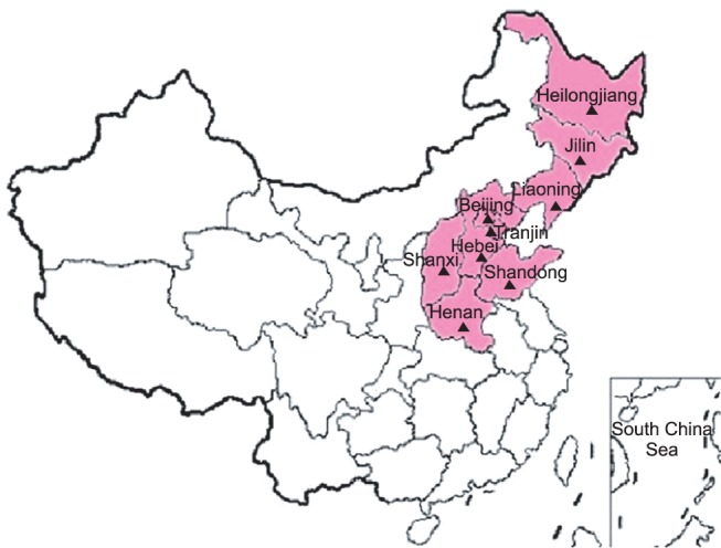
Regions of pseudorabies virus (PRV) outbreak in China. The provinces or autonomous cities affected are indicated in pink. Regions marked with black triangles indicate areas showing positive PRV results as detected by enzyme-linked immunosorbent assay and polymerase chain reaction analyses.
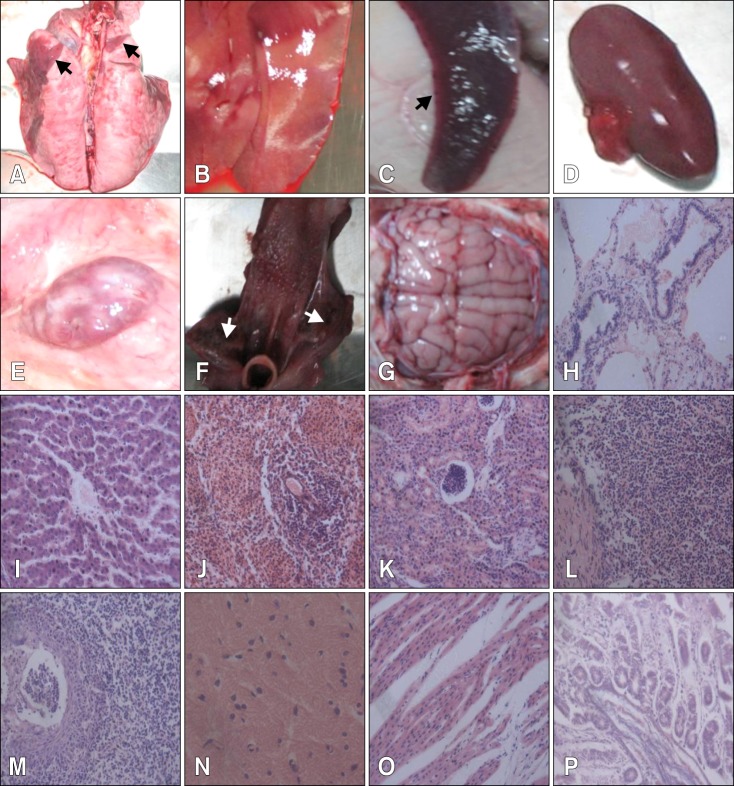
Severe damage to multiple organs in experimental, infected piglets by postmortem and histopathological examinations. (A) Lung necrosis (arrows). (B) Liver with yellowish white spots indicating necrosis or hemorrhage. (C) Spleen infarct (arrow). (D) Kidney with bleeding spots. (E) Hemorrhagic lymph node. (F) Tonsil necrosis (arrows). (G) Slight encephalic edema. (H) Alveolar ducts and terminal bronchiolar cavities filled with cellular and serous exudates. (I) Swelling and degeneration of liver cells. (J) Splenic cord with unclear structure and reduced lymphocytes. (K) Swelling and disintegration of epithelial cells. (L) Reduced lymphoid nodules with irregular structures. (M) Epithelial cells filled with eosinophilic intranuclear inclusions. (N) Glial cells with neurons. (O) Breakage and disintegration of myocardial fibers. (P) Midgut gland atrophy. H&E stain, ×400.
References
-
- Cramer SD, Campbell GA, Njaa BL, Morgan SE, Smith SK, 2nd, McLin WR, 4rd, Brodersen BW, Wise AG, Scherba G, Langohr IM, Maes RK. Pseudorabies virus infection in Oklahoma hunting dogs. J Vet Diagn Invest. 2011;23:915–923. - PubMed
-
- Marcaccini A, Peña ML, Quiroga MI, Bermúdez R, Nieto JM, Alemañ N. Pseudorabies virus infection in mink: a host-specific pathogenesis. Vet Immunol Immunopathol. 2008;124:264–273. - PubMed
-
- Müller T, Hahn EC, Tottewitz F, Kramer M, Klupp BG, Mettenleiter TC, Freuling C. Pseudorabies virus in wild swine: a global perspective. Arch Virol. 2011;156:1691–1705. - PubMed
Publication types
MeSH terms
LinkOut - more resources
Full Text Sources
Other Literature Sources
