Review
. 2013 Jul 2;4(4):e00413-13.
doi: 10.1128/mBio.00413-13.

Envelope protein dynamics in paramyxovirus entry

Affiliations
Review

Envelope protein dynamics in paramyxovirus entry

Philippe Plattet et al. mBio. .

Abstract

Paramyxoviruses include major pathogens with significant global health and economic impact. This large family of enveloped RNA viruses infects cells by employing two surface glycoproteins that tightly cooperate to fuse their lipid envelopes with the target cell plasma membrane, an attachment and a fusion (F) protein. Membrane fusion is believed to depend on receptor-induced conformational changes within the attachment protein that lead to the activation and subsequent refolding of F. While structural and mechanistic studies have considerably advanced our insight into paramyxovirus cell adhesion and the structural basis of F refolding, how precisely the attachment protein links receptor engagement to F triggering remained poorly understood. Recent reports based on work with several paramyxovirus family members have transformed our understanding of the triggering mechanism of the membrane fusion machinery. Here, we review these recent findings, which (i) offer a broader mechanistic understanding of the paramyxovirus cell entry system, (ii) illuminate key similarities and differences between entry strategies of different paramyxovirus family members, and (iii) suggest new strategies for the development of novel therapeutics.


Figures

FIG 1
FIG 1
(A) Side views of PIV5 HN (PDB code, 1Z4Z), NiV G bound to ephrinB2 (PDB code, 3D12), MeV H monomer bound to SLAM (PDB code, 3ALZ) and MeV H monomer bound to nectin-4 (PDB code, 4GJT). Beta-propeller structures of paramyxovirus attachment protein head domains are shown in red, whereas the proteinaceous receptors are shown in cyan. (B) Top views of the MeV H dimer (left) and tetramer bound to SLAM (right) (PDB code, 3ALZ). In the MeV H tetramer, one dimer of dimers is shown in red, and the other is in green.
FIG 2
FIG 2
Side views of two forms of MeV-H tetramer complexed with SLAM (cyan). One MeV-H dimer is shown in red, and the other is in green. The tetramers assume two distinct conformations (A [PDB code, 3ALZ] and B [PDB code, 3ALX]).
FIG 3
FIG 3
(A) Structure of the soluble NDV HN tetramer ectodomain in the heads-down conformation (PDB code, 3T1E). In this form, the two lower heads of each dimer interact with the stalk (in the 4HB conformation). The four connectors linking the top of the stalk to the four different heads are not present in the X-ray structures. (B) Structure of the soluble PIV5 HN stalk domain, consisting of an upper “straight” region and a lower “supercoiled” region (PDB code, 3TSI). (C) Putative pre-receptor-binding conformation of membrane-embedded full-length PIV5/NDV HN tetramers.
FIG 4
FIG 4
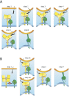
Models of receptor-induced conformational changes of the paramyxovirus attachment protein tetramer that are considered to result in F triggering. (A) Proposed model for morbilliviruses and henipaviruses. Prior to receptor binding, H/G tetrameric proteins associate with F trimers (step 1). The attachment protein then binds to its cognate cell surface receptor, which, in turn, leads to the tethering of the two opposing lipid bilayers (viral envelope and host cell plasma membrane), creating a fusion-competent microenvironment (step 2). As a result of receptor binding by the H/G head domains, the tetrameric heads undergo a “sliding” movement (step 3). Consequently, the central section of the tetrameric stalk unwinds/unfolds (step 4). Since the H/G stalk section that undergoes conformational changes is in short-range contact with F, the preassembled hetero-oligomeric H/G-F fusion complexes dissociate (step 5), and the destabilized or liberated F trimers subsequently undergo irreversible structural rearrangements (represented by F reaching the prehairpin structural intermediates; step 6). F conformational changes progress to the postfusion state, which ultimately leads to fusion pore formation (not shown). (B) Proposed model for rubulaviruses, avulaviruses, and respiroviruses. Prior to receptor binding, HN tetramers assume a four-heads-down conformation that (i) prevents functional hetero-oligomeric assembly with F trimers and (ii) stabilizes the stalk domain in a pre-receptor-binding state (step 1). Upon receptor binding, the four HN heads move up, which in turn enables F trimers to contact HN (step 2a). The HN stalk domain then refolds into the post-receptor-binding state (trigger-competent stalk domain) (step 3). Alternatively, receptor-induced movements may coincide with immediate stalks’ rearrangements and may be followed by F binding (step 2b). Common to both models, this post-receptor-binding trigger-competent central stalk section then destabilizes F trimers (step 3). As in panel A, these F trimers undergo irreversible structural rearrangements that lead to fusion pore formation (step 4). Prefusion F trimers are based on a high-resolution structural model that was morphed into a lower-resolution image using the Sculptor package. For the sake of clarity, only one receptor unit is represented in the cartoon. The host plasma membrane is in orange, and the viral envelope is in blue.

References

    1. Chua KB, Bellini WJ, Rota PA, Harcourt BH, Tamin A, Lam SK, Ksiazek TG, Rollin PE, Zaki SR, Shieh W, Goldsmith CS, Gubler DJ, Roehrig JT, Eaton B, Gould AR, Olson J, Field H, Daniels P, Ling AE, Peters CJ, Anderson LJ, Mahy BW. 2000. Nipah virus: a recently emergent deadly paramyxovirus. Science 288:1432–1435 - PubMed
    1. Murray K, Selleck P, Hooper P, Hyatt A, Gould A, Gleeson L, Westbury H, Hiley L, Selvey L, Rodwell B, Ketterer P. 1995. A morbillivirus that caused fatal disease in horses and humans. Science 268:94–97 - PubMed
    1. Lamb RA, Parks GD. 2007. Paramyxoviridae: the viruses and their replication, p 1449–1496 In Knipe DM, Howley PM, Fields virology, 5th ed. Lippincott Williams & Wilkins, Philadelphia, PA.
    1. Plemper RK. 2011. Cell entry of enveloped viruses. Curr. Opin. Virol. 1:92–100 - PMC - PubMed
    1. Yin HS, Wen X, Paterson RG, Lamb RA, Jardetzky TS. 2006. Structure of the parainfluenza virus 5 F protein in its metastable, prefusion conformation. Nature 439:38–44 - PMC - PubMed

Publication types

Substances