

Proteomic analysis of the SH2 domain-containing leukocyte protein of 76 kDa (SLP76) interactome in resting and activated primary mast cells [corrected]
- PMID: 23820730
- PMCID: PMC3790297
- DOI: 10.1074/mcp.M112.025908
Proteomic analysis of the SH2 domain-containing leukocyte protein of 76 kDa (SLP76) interactome in resting and activated primary mast cells [corrected]
Erratum in
- Mol Cell Proteomics. 2013 Feb ;13(2):678
-
Proteomic Analysis of the SH2 Domain-containing Leukocyte Protein of 76 kDa (SLP76) Interactome in Resting and Activated Primary Mast Cells.Mol Cell Proteomics. 2014 Feb;13(2):678. doi: 10.1074/mcp.A112.025908. Epub 2020 Oct 1. Mol Cell Proteomics. 2014. PMID: 33498147 Free PMC article. No abstract available.
Abstract
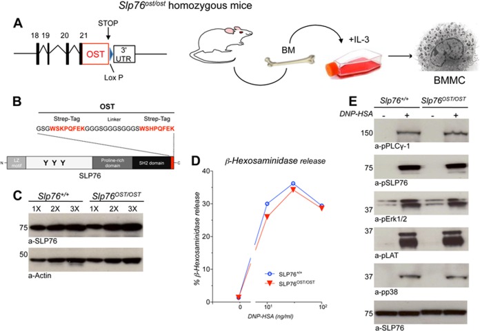
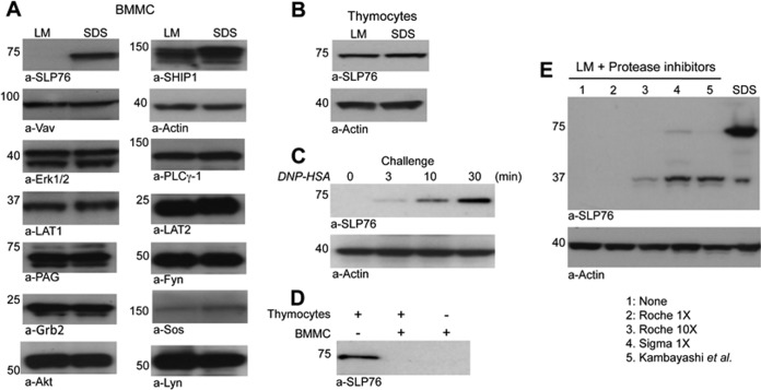
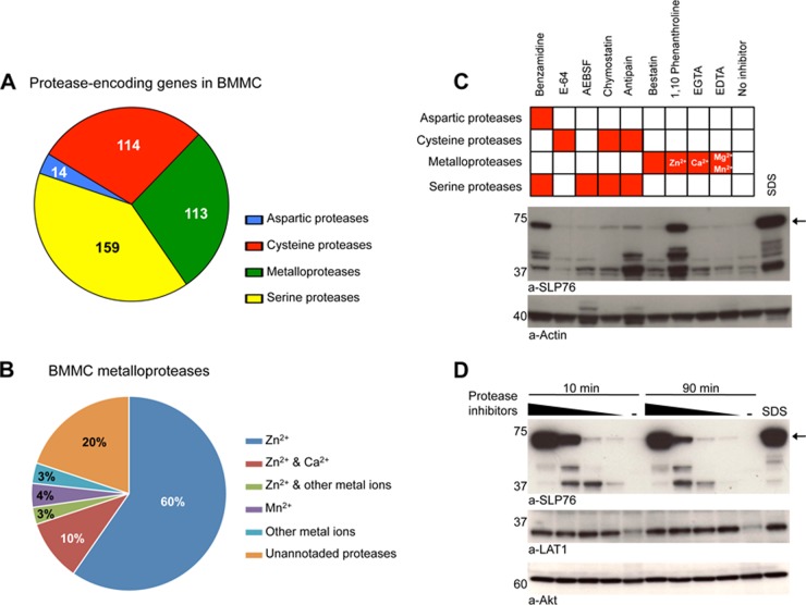
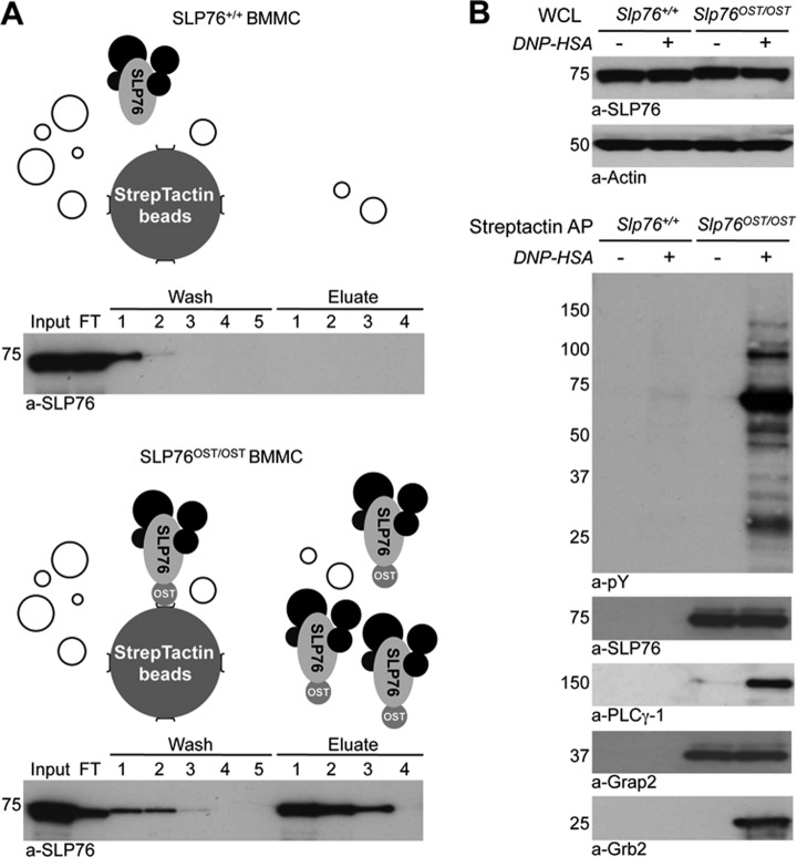
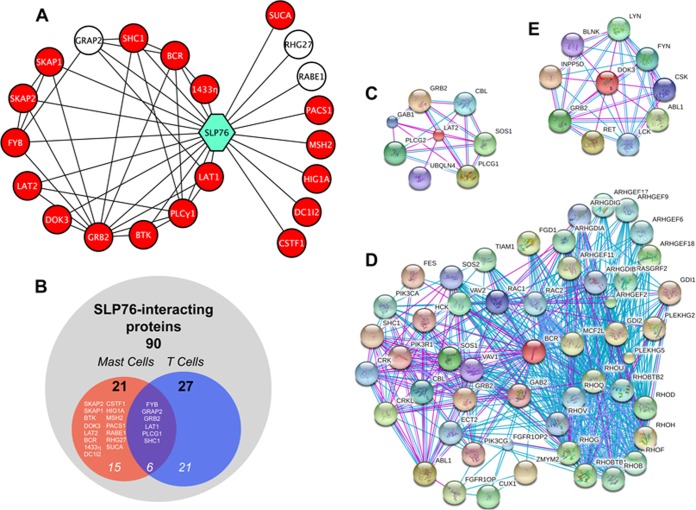
We report the first proteomic analysis of the SLP76 interactome in resting and activated primary mouse mast cells. This was made possible by a novel genetic approach used for the first time here. It consists in generating knock-in mice that express signaling molecules bearing a C-terminal tag that has a high affinity for a streptavidin analog. Tagged molecules can be used as molecular baits to affinity-purify the molecular complex in which they are engaged, which can then be studied by mass spectrometry. We examined first SLP76 because, although this cytosolic adapter is critical for both T cell and mast cell activation, its role is well known in T cells but not in mast cells. Tagged SLP76 was expressed in physiological amounts and fully functional in mast cells. We unexpectedly found that SLP76 is exquisitely sensitive to mast cell granular proteases, that Zn(2+)-dependent metalloproteases are especially abundant in mast cells and that they were responsible for SLP76 degradation. Adding a Zn(2+) chelator fully protected SLP76 in mast cell lysates, thereby enabling an efficient affinity-purification of this adapter with its partners. Label-free quantitative mass spectrometry analysis of affinity-purified SLP76 interactomes uncovered both partners already described in T cells and novel partners seen in mast cells only. Noticeably, molecules inducibly recruited in both cell types primarily concur to activation signals, whereas molecules recruited in activated mast cells only are mostly associated with inhibition signals. The transmembrane adapter LAT2, and the serine/threonine kinase with an exchange factor activity Bcr were the most recruited molecules. Biochemical and functional validations established the unexpected finding that Bcr is recruited by SLP76 and positively regulates antigen-induced mast cell activation. Knock-in mice expressing tagged molecules with a normal tissue distribution and expression therefore provide potent novel tools to investigate signalosomes and to uncover novel signaling molecules in mast cells.
Figures

Similar articles
-
Protein tyrosine phosphatase epsilon is a negative regulator of FcepsilonRI-mediated mast cell responses.Scand J Immunol. 2009 May;69(5):401-11. doi: 10.1111/j.1365-3083.2009.02235.x. Epub 2008 Feb 6. Scand J Immunol. 2009. PMID: 19508371
-
Serine Phosphorylation of SLP76 Is Dispensable for T Cell Development but Modulates Helper T Cell Function.PLoS One. 2017 Jan 20;12(1):e0170396. doi: 10.1371/journal.pone.0170396. eCollection 2017. PLoS One. 2017. PMID: 28107427 Free PMC article.
-
Independent and cooperative roles of adaptor molecules in proximal signaling during FcepsilonRI-mediated mast cell activation.Mol Cell Biol. 2010 Sep;30(17):4188-96. doi: 10.1128/MCB.00305-10. Epub 2010 Jul 6. Mol Cell Biol. 2010. PMID: 20606011 Free PMC article.
-
A perspective: regulation of IgE receptor-mediated mast cell responses by a LAT-organized plasma membrane-localized signaling complex.Int Arch Allergy Immunol. 2001 Jan-Mar;124(1-3):137-41. doi: 10.1159/000053692. Int Arch Allergy Immunol. 2001. PMID: 11306950 Review.
-
Cooperation of adapter molecules in proximal signaling cascades during allergic inflammation.Immunol Rev. 2009 Nov;232(1):99-114. doi: 10.1111/j.1600-065X.2009.00825.x. Immunol Rev. 2009. PMID: 19909359 Review.
Cited by
-
Identification of key genes in salivary gland in Sjögren's syndrome complicated with Hashimoto thyroiditis: Common pathogenesis and potential diagnostic markers.Medicine (Baltimore). 2023 Sep 29;102(39):e35188. doi: 10.1097/MD.0000000000035188. Medicine (Baltimore). 2023. PMID: 37773833 Free PMC article.
-
Proteome analysis of mast cell releasates reveals a role for chymase in the regulation of coagulation factor XIIIA levels via proteolytic degradation.J Allergy Clin Immunol. 2017 Jan;139(1):323-334. doi: 10.1016/j.jaci.2016.03.051. Epub 2016 May 13. J Allergy Clin Immunol. 2017. PMID: 27302551 Free PMC article.
-
Automated inference of Boolean models from molecular interaction maps using CaSQ.Bioinformatics. 2020 Aug 15;36(16):4473-4482. doi: 10.1093/bioinformatics/btaa484. Bioinformatics. 2020. PMID: 32403123 Free PMC article.
-
Butyrate Selectively Targets Super-Enhancers and Transcriptional Networks Associated with Human Mast Cell Function.Eur J Immunol. 2025 Jun;55(6):e51680. doi: 10.1002/eji.202451680. Eur J Immunol. 2025. PMID: 40498295 Free PMC article.
-
Decoding communication patterns of the innate immune system by quantitative proteomics.J Leukoc Biol. 2019 Dec;106(6):1221-1232. doi: 10.1002/JLB.2RI0919-302R. Epub 2019 Sep 26. J Leukoc Biol. 2019. PMID: 31556465 Free PMC article. Review.
References
-
- Kinet J. P. (1999) The high-affinity IgE receptor (Fc epsilon RI): from physiology to pathology. Annu. Rev. Immunol. 17, 931–972 - PubMed
-
- Reth M. (1989) Antigen receptor tail clue. Nature 338, 383–384 - PubMed
-
- Blank U., Ra C., Miller L., White K., Metzger H., Kinet J. P. (1989) Complete structure and expression in transfected cells of high affinity IgE receptor. Nature 337, 187–189 - PubMed
-
- Turner H., Kinet J. P. (1999) Signalling through the high-affinity IgE receptor Fc epsilonRI. Nature 402, B24–B30 - PubMed
Publication types
MeSH terms
Substances
LinkOut - more resources
Full Text Sources
Other Literature Sources
Miscellaneous
