. 2013 Oct;21(10):1909-18.
doi: 10.1038/mt.2013.152. Epub 2013 Jul 3.

Silencing mutant ATXN3 expression resolves molecular phenotypes in SCA3 transgenic mice

Affiliations

Silencing mutant ATXN3 expression resolves molecular phenotypes in SCA3 transgenic mice

Edgardo Rodríguez-Lebrón et al. Mol Ther. 2013 Oct.

Erratum in

  • Mol Ther. 2014 Apr;22(4):891. Costa, Maria doCarmo [corrected to Costa, Maria do Carmo]

Abstract

Spinocerebellar ataxia type 3 (SCA3) is a neurodegenerative disease caused by a polyglutamine expansion in the deubiquitinating enzyme, Ataxin-3. Currently, there are no effective treatments for this fatal disorder but studies support the hypothesis that reducing mutant Ataxin-3 protein levels might reverse or halt the progression of disease in SCA3. Here, we sought to modulate ATXN3 expression in vivo using RNA interference. We developed artificial microRNA mimics targeting the 3'-untranslated region (3'UTR) of human ATXN3 and then used recombinant adeno-associated virus to deliver them to the cerebellum of transgenic mice expressing the full human disease gene (SCA3/MJD84.2 mice). Anti-ATXN3 microRNA mimics effectively suppressed human ATXN3 expression in SCA3/MJD84.2 mice. Short-term treatment cleared the abnormal nuclear accumulation of mutant Ataxin-3 throughout the transduced SCA3/MJD84.2 cerebellum. Analysis also revealed changes in the steady-state levels of specific microRNAs in the cerebellum of SCA3/MJD84.2 mice, a previously uncharacterized molecular phenotype of SCA3 that appears to be dependent on mutant Ataxin-3 expression. Our findings support the preclinical development of molecular therapies aimed at halting the expression of ATXN3 as a viable approach to SCA3 and point to microRNA deregulation as a potential surrogate marker of SCA3 pathogenesis.


Figures

Figure 1
Figure 1
Targeting human ATXN3 expression with RNA interference. (a) The 3′UTR of ATXN3 was scanned for siRNA target sites and the top five candidates, numbered by their position with respect to the ATXN3 UAA stop codon (RefSeq: NM_004993), were experimentally validated. (b) A representative western blot analysis of endogenous Ataxin-3 expression following delivery of siRNAs into HEK293 cells. Compared with untreated (null) cells or those receiving a scramble siRNA control (miss), ectopic delivery of anti-ATXN3 siRNAs (148, 149, 316, 501, and 673) led to a marked reduction in the levels of Ataxin-3 protein 72-hours post-transfection. The results from four biological replicates are graphed to the right of the blot. Ataxin-3 protein levels were normalized to those of α-tubulin (Tub). (c) Scheme of miR-Atx3-148 miRNA mimic. siRNA sequences were embedded (grayscale-shaded region) in a miRNA-like RNA scaffold (black nucleotides) that mimics the human miRNA-124. The U6 snRNA promoter was used to drive intracellular expression of the miRNA mimics. Closed and opened triangles indicate the predicted Drosha and Dicer cleavage sites, respectively. (d) Northern blot analysis was used to compare the intracellular production and processing of the anti-ATXN3 miRNA mimic (miR-Atx3-148) to that of a “first-generation” short-hairpin RNA design (sh-Atx3-148), a miR-Atx3-148 construct lacking the basal stem sequence (-basal stem) and a miR-Atx3-148 construct lacking the loop sequence (-loop seq). Accumulated precursor RNA molecules were detected in the sh-Atx3-148 (black arrowhead) and -basal stem (white arrowhead) lanes using a probe against the guide strand sequence. Approximately equal levels of fully processed guide-strand were detected (bottom arrow) in all samples but the -loop seq sample. RNA levels were normalized to those of the 5S small noncoding RNA (asterisk). The molecular marker (mm) is shown in the first lane. (e) Analysis ofATXN3 mRNA levels following transient expression of anti-ATXN3 miRNA mimics in HEK293 cells. miR-Atx3-148 and miR-Atx3-149 mediated effective gene silencing of endogenous human ATXN3 mRNA when compared with untreated (null) or scramble-control (miss) treated cells. Error bars: ±SD.
Figure 2
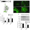
Figure 2
rAAV2/1-mediated delivery of miR-Atx3-148 to the cerebellum of SCA3/MJD84.2 mice leads to gene silencing of human mutant Ataxin-3. (a) The U6-miRNA expression cassette was cloned into a recombinant AAV expression vector upstream of a CMV-driven hrGFP expression cassette. rAAV2/1 virus was delivered into the deep cerebellar nuclei (DCN) of SCA3/MJD84.2 transgenic mice. Purkinje neurons in the cerebellar cortex, which project their axons to the DCN, are also transduced by DCN-delivered rAAV2/1 via retrograde transport. (be) Cerebellar coronal sections were obtained from injected mice 8 weeks post-surgery. Confocal microscopy analysis revealed strong hrGFP expression throughout neurons in the DCN (b, c) and in cerebellar Purkinje neurons (d, e).Scale bar in (b) and (d) = 200 µm. Scale bar in (c) and (e) = 50 µm. (f) Mutant human ATXN3 mRNA levels were suppressed following delivery of the rAAV2/1 miR-Atx3-148 virus. In contrast, the steady state levels of mouse Atxn3 and Calbindin-1 mRNAs remained unchanged in the presence of rAAV-miR-Atx3-148 virus. (g) Cerebellar mutant Ataxin-3 protein expression was confirmed by western blot using the 1H9 antibody. This antibody recognizes human mutant Ataxin-3 (top band) and endogenous mouse ataxin-3 (lower band). Four control-injected (lanes 1–4) and four rAAV-miR-148-injected (lanes 5–8) SCA3/84.2 mice were compared. As a group, SCA3/84.2 mice injected with rAAV-miR-148 showed a significant reduction (~34%) in mutant Ataxin-3 levels when compared to control injected mice. Gapdh levels were used to normalize for protein loading.*P < 0.05, unpaired t test.
Figure 3
Figure 3
miR-Atx3-148 activity resolves neuronal nuclear accumulation of mutant Atx3 in SCA3/84.2 cerebellum. (a) Mutant human Ataxin-3 expression was analyzed immunohistochemically using the 1H9 monoclonal antibody against human Ataxin-3 (red color). hrGFP expression was evident in neurons throughout the ipsilateral DCN (a) but absent in the contralateral, uninjected DCN where only a few hrGFP-positive tracks could be observed (b). As expected, hrGFP-positive areasin the rAAV2/1-miR-Atx3-148 injected DCN of SCA3/MJD84.2 mice were almost completely devoid of mutant Ataxin-3 signal (c, ipsilateral) whereas intense neuronal nuclear mutant Ataxin-3 staining was detected throughout the contralateral DCN of SCA3/MJD84.2 mice (d, contralateral). This mutant Ataxin-3 “exclusion zone” is highlighted in the merged image panels (e) and (f). (gi) In contrast, mutant Ataxin-3 was still clearly present inthe nuclei of DCN neurons expressing the control hrGFP-only vector. Arrow in (i) points to DCN neurons transduced with rAAV-CMV-hrGFP control vector (hrGFP-positive) while the arrowhead points to a neighboring hrGFP-negative DCN neuron. Notice the similar levels of nuclear mutant Ataxin-3 expression between neurons transduced with the control vector and untransduced DCN neurons, as seen in the merged image (i). (jo) hrGFP signal in the cerebellar cortex of injected SCA3/MJD84.2 mice predominantly localized to Purkinje neurons. The anti-Ataxin-31H9 signal (red) was absent in Purkinje neurons expressing rAAV-miR-Atx3-148 (jl, left side of lobule) but easily detected in the nuclei and processes of untransduced Purkinje neurons (jl, right side of lobule). In comparison, nuclear mutant Ataxin-3 signal was observed in Purkinje neurons expressing the control rAAV2/1-CMV-hrGFP vector (mo). Scale bar in (af) = 200 µm. Scale bar in (go) = 50 µm.
Figure 4
Figure 4
Silencing mutant Ataxin-3 leads to a partial normalization of endogenous miRNA expression in SCA3/84.2 mice. Global miRNA expression profiles for 2-month (a) and 6-month (b) old SCA3/84.2 and littermate control mice (n = 3 per group). The volcano plot shows the magnitude (x-axis, log2 fold change) and the statistical significance (y-axis, -log10 P value) of the calculated difference in miRNA expression. miRNAs with a significant (P < 0.05, above horizontal dash line) fold increase (≥1.2, upper right quadrant) or decrease (≥1.2, upper left quadrant) in steady-state levels in SCA3/84.2 mice relative to age-matched littermate controls are represented by black filled circles. Grey-filled circles represent expressed cerebellar miRNAs with similar expression levels in SCA3/84.2 relative to age-matched littermate controls. At 2-months of age, 10 different miRNAs (listed in the “Results” section) significantly differed between SCA3/84.2 and littermate control mice. By 6-months of age, only the steady-state levels of miR-181a were significantly altered between transgenic and control mice. (c) A subset of miRNAs (miR-9, -16, -124, -128 and -30c) that displayed similar steady-state levels in untreated SCA3/84.2 mice relative to littermate controls (e.g., grey-filled circles) was used to detect potential adverse effects of sustained miR-Atx3-148 expression on the endogenous mouse miRNA pathway. Following 8 weeks of miR-Atx3-148 expression in SCA3/84.2 cerebellar neurons, there was no significant difference in the levels of miR-9, -16, -124, -128, or -30c in transduced SCA3/84.2 mice compared with untreated (SCA3 TG) or control injected (SCA3 TG-Ctrl) transgenic mice. (d) Delivery of miR-Atx3-148 expression, however, appeared to partially normalize steady-state levels of miR-181a and miR-674, two of the miRNAs with altered steady-state levels in the cerebella of untreated SCA3/84.2 mice. The graph shows steady-state levels of each miRNA in SCA3/84.2 mice expressing miR-Atx3-148 (SCA3 TG-148, black bars) relative to control treated SCA3/84.2 mice (SCA3 TG-Ctrl, white bars). Since miR-181a levels were increased by ~twofold in untreated SCA3/84.2 mouse cerebellum (see panels a and b), a reduction in its levels following suppression of human ATXN3 suggests a partial restoration to control levels. *P < 0.05, unpaired t test.

References

    1. Costa Mdo C, Paulson HL. Toward understanding Machado-Joseph disease. Prog Neurobiol. 2012;97:239–257. - PMC - PubMed
    1. Matos CA, de Macedo-Ribeiro S, Carvalho AL. Polyglutamine diseases: the special case of ataxin-3 and Machado-Joseph disease. Prog Neurobiol. 2011;95:26–48. - PubMed
    1. Durr A. Autosomal dominant cerebellar ataxias: polyglutamine expansions and beyond. Lancet Neurol. 2010;9:885–894. - PubMed
    1. Boy J, Schmidt T, Wolburg H, Mack A, Nuber S, Böttcher M, et al. Reversibility of symptoms in a conditional mouse model of spinocerebellar ataxia type 3. Hum Mol Genet. 2009;18:4282–4295. - PubMed
    1. Yamamoto A, Lucas JJ, Hen R. Reversal of neuropathology and motor dysfunction in a conditional model of Huntington's disease. Cell. 2000;101:57–66. - PubMed

Publication types

MeSH terms