Review
. 2013 Jul;7 Suppl 1(Suppl 1):S68-76.
doi: 10.1007/s12105-013-0454-z. Epub 2013 Jul 3.

Carcinoma ex pleomorphic adenoma, with particular emphasis on early lesions

Affiliations
Review

Carcinoma ex pleomorphic adenoma, with particular emphasis on early lesions

Silvana Di Palma. Head Neck Pathol. 2013 Jul.

Abstract

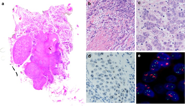
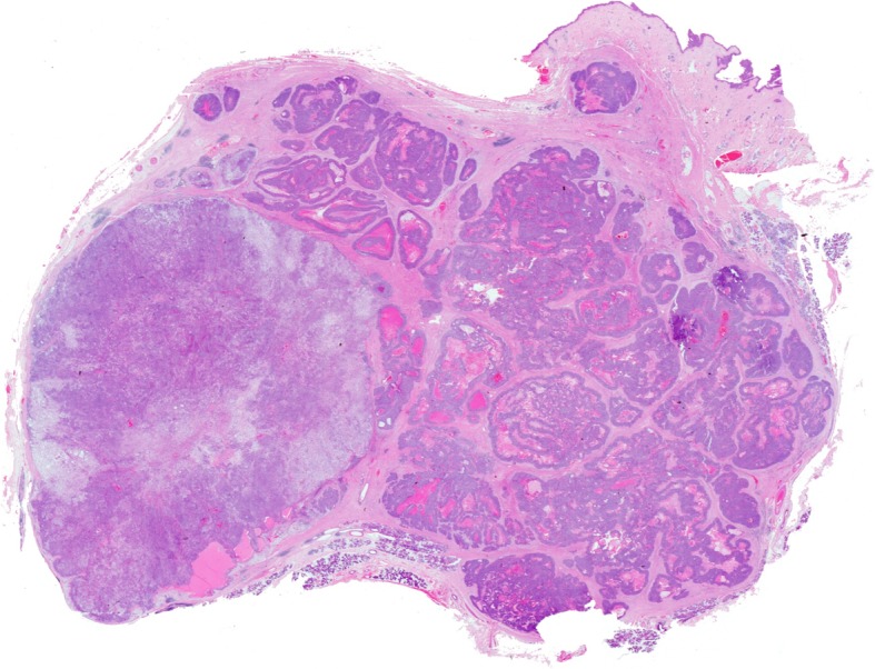
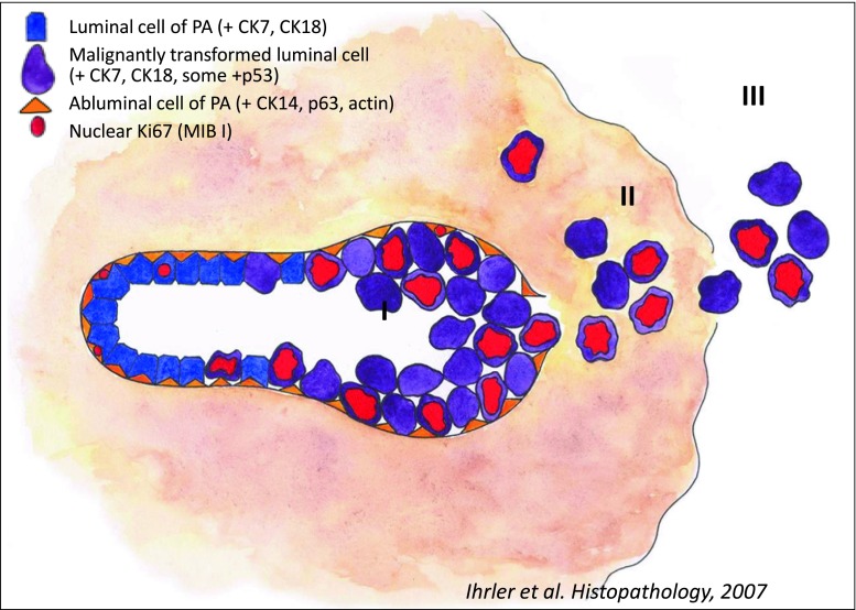
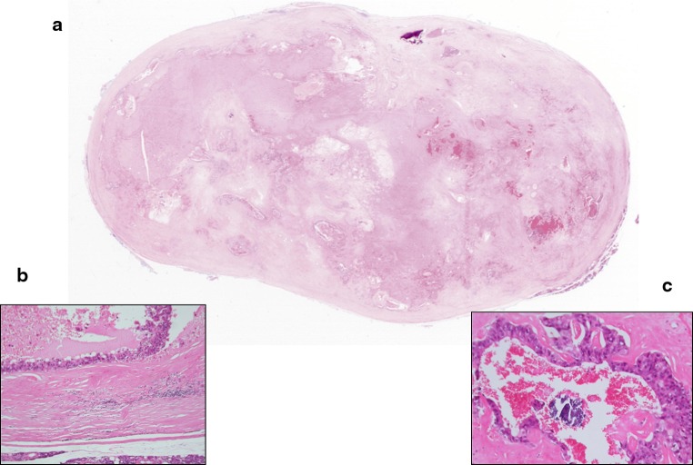
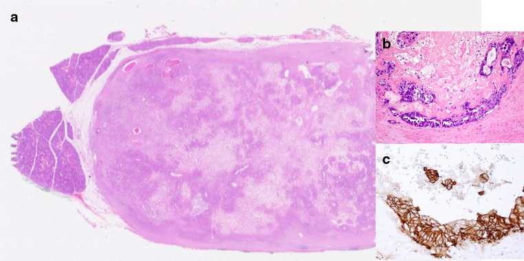
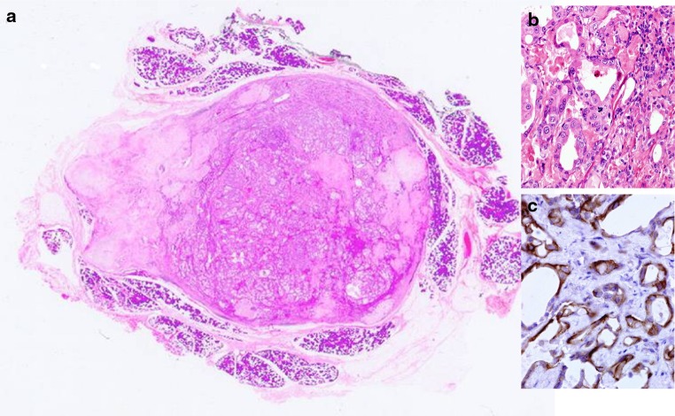
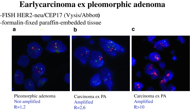
Carcinoma ex pleomorphic adenoma (CXPA) is a broad category of carcinomas of the salivary glands which includes at least 2 clinically relevant categories; one is referred here as early CXPA (ECXPA), the other as widely invasive CXPA. The former includes several histological patterns ranging from non-invasive/in situ/intraductal/intratubular, early invasive/extratubular/intracapsular and extracapsular (up to 6 mm). The latter includes any CXPA with invasion of >6 mm. The clinical behaviour of ECXPA is not aggressive and tends to overlap that of a pleomorphic adenoma (PA) which makes the histological report of carcinoma contradictory. These early malignant changes in PA are known since the 1970s but it has been the use of immunohistochemical and molecular genetic analysis for HER-2 and TP53 gene in the last decade that has clarified the genuine malignant nature of the cells. HER-2 and TP53 gene and protein are involved in the early stages of malignant transformation of PA. Moreover the immunohistochemical over-expression HER-2, p53 protein and Mib-1 proliferation marker may be useful markers to identify malignant areas in PA.


Figures

Fig. 1
Fig. 1
Whole mounted section of carcinoma ex Pleomorphic adenoma of parotid gland with perineural infiltration (b) and highly pleomorphic cells with numerous mitoses (c). The maternal PA shows sclerosis and calcifications. HER-2/neu gene amplification detected by Silver (d) and fluorescent (e) in situ hybridization
Fig. 2
Fig. 2
Whole mounted section of myoepithelial carcinoma (right) arising in Pleomorphic Adenoma (left) of parotid. Note the multinodular growth pattern with infiltration of the dermis
Fig. 3
Fig. 3
Diagram showing progression of carcinoma ex pleomorphic adenoma. I Pre-existing tubules of Pleomorphic Adenoma partly replaced by malignant epithelial cells. II Rupture of basal membrane with extratubular extension but still confined within the capsule of PA. III Extracapsular spread with infiltration of surrounding tissues. Courtesy by Dr. S. Ihrler
Fig. 4
Fig. 4
Whole mounted section of Early Carcinoma Ex Pleomorphic adenoma (ECXPA). The nodule is well circumscribed and there is no extension into surrounding tissues. Calcifications and haemorrhage is seen. The viable areas consist of ducts lined by atypical cells b with early cribriform architecture c
Fig. 5
Fig. 5
Another example of ECXPA where the atypical cells show strong membrane staining for HER-2/neu (score 3+). (Immunohistochemical staining, original magnification ×360)
Fig. 6
Fig. 6
ECXPA. Whole mounted section a. Atypical epithelial proliferation replacing ducts of PA b. Moderate membrane staining for HER-2/neu (score 2+) in atypical cells of carcinoma ex pleomorphic adenoma. Note absence of staining in benign component. c (immunohistochemical staining, original magnification ×80)
Fig. 7
Fig. 7
ECXPA. a No amplification of HER-2/neu gene in PA, b and c low and high level of amplification, with 2.6 and >10 mean number of gene copies per nucleus (FISH)

Similar articles

Cited by

References

    1. Gnepp DR, Brandwein-Gensler MS, El-Naggar AK, Nagao T. Carcinoma ex pleomorphic adenoma. In: Barnes EL, Everson JW, El-Naggar AK, editors.WHO classification of tumors, vol. 9. 2005. p.242–3.
    1. Auclair PL, Ellis GL. Atypical features in salivary gland mixed tumors: their relationship to malignant transformation. Mod Pathol. 1996;9:652–657. - PubMed
    1. Skálová A, Altemani A, Di Palma S, Simpson R, Hostička L, Andrle P, Laco J, Toner M, Vozmitsel M, Szakacs S, Kazakov D, Kinkor Z, Michal M. Pleomorphic adenoma of the salivary glands with intravascular tumor deposits, A diagnostic pitfall. Am J Surg Pathol. 2012;36:1674–1682. doi: 10.1097/PAS.0b013e3182690afe. - DOI - PubMed
    1. Di Palma S, Skálová A, Vanìèek T, Simpson RH, Stárek I, Leivo I. Non-invasive (intracapsular) carcinoma ex pleomorphic adenoma: recognition of focal carcinoma by HER2/neu and MIB1 immunohistochemistry. Histopathology. 2005;46:144–152. doi: 10.1111/j.1365-2559.2005.02058.x. - DOI - PubMed
    1. Wenig BM, Hitchcock CL, Ellis GL, Gnepp DR. Metastasizing mixed tumour of salivary glands: a clinicopathologic and flow cytometric analysis. Am J Surg Pathol. 1992;16:845–858. doi: 10.1097/00000478-199209000-00003. - DOI - PubMed

MeSH terms

Substances