"Dedifferentiation" and high-grade transformation in salivary gland carcinomas
- PMID: 23821210
- PMCID: PMC3712099
- DOI: 10.1007/s12105-013-0458-8
"Dedifferentiation" and high-grade transformation in salivary gland carcinomas
Abstract
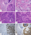
"Dedifferentiation" and/or high-grade transformation (HGT) has been described in a variety of salivary gland carcinomas, including acinic cell carcinoma, adenoid cystic carcinoma, epithelial-myoepithelial carcinoma, polymorphous low-grade adenocarcinoma, myoepithelial carcinoma, low-grade mucoepidermoid carcinoma and hyalinizing clear cell carcinoma, although the phenomenon is a rare event. Recent authors tend to preferably use the term HGT instead of "dedifferentiation" in these cases. HGT-tumors are composed of conventional carcinomas juxtaposed with areas of HG morphology, usually either poorly differentiated adenocarcinoma or "undifferentiated" carcinoma, in which the original line of differentiation is no longer evident. The HG component is generally composed of solid nests, sometimes occurring in cribriform pattern of anaplastic cells with large vesicular pleomorphic nuclei, prominent nucleoli and abundant cytoplasm. Frequent mitoses and extensive necrosis is evident. The Ki-67 labeling index is consistently higher in the HG component. p53 abnormalities have been demonstrated in the transformed component in a few examples, but the frequency varies by the histologic type. HER-2/neu overexpression and/or gene amplification is considerably exceptional. The molecular-genetic mechanisms responsible for the pathway of HGT in salivary gland carcinomas largely still remain to be elucidated. Salivary gland carcinomas with HGT have been shown to be more aggressive than conventional carcinomas with a poorer prognosis, accompanied by higher local recurrence rate and propensity for cervical lymph node metastasis, suggesting the need for wider resection and neck dissection.
Figures


Similar articles
-
High-grade Transformation/Dedifferentiation in Salivary Gland Carcinomas: Occurrence Across Subtypes and Clinical Significance.Adv Anat Pathol. 2021 May 1;28(3):107-118. doi: 10.1097/PAP.0000000000000298. Adv Anat Pathol. 2021. PMID: 33825717 Review.
-
High-grade transformation/dedifferentiation of an adenoid cystic carcinoma of the minor salivary gland to myoepithelial carcinoma.Pathol Int. 2018 Feb;68(2):133-138. doi: 10.1111/pin.12624. Epub 2017 Dec 29. Pathol Int. 2018. PMID: 29287310
-
Metastatic adenoid cystic carcinoma with high-grade transformation ("dedifferentiation") in pleural effusion and neck lymph node: A diagnostic challenge on cytology?Diagn Cytopathol. 2020 Jul;48(7):679-683. doi: 10.1002/dc.24431. Epub 2020 Apr 9. Diagn Cytopathol. 2020. PMID: 32271503
-
Fine needle aspiration of salivary gland carcinomas with high-grade transformation: A multi-institutional study of 22 cases and review of the literature.Cancer Cytopathol. 2021 Apr;129(4):318-325. doi: 10.1002/cncy.22388. Epub 2020 Nov 19. Cancer Cytopathol. 2021. PMID: 33211402 Free PMC article. Review.
-
Adenoid cystic carcinoma with high-grade transformation: a report of 11 cases and a review of the literature.Am J Surg Pathol. 2007 Nov;31(11):1683-94. doi: 10.1097/PAS.0b013e3180dc928c. Am J Surg Pathol. 2007. PMID: 18059225 Review.
Cited by
-
Insight into the Evolving Concepts on the Origin of Salivary Gland Neoplasms.J Int Oral Health. 2015;7(Suppl 1):i-ii. J Int Oral Health. 2015. PMID: 26225117 Free PMC article. No abstract available.
-
High-Grade Transformation in Adenoid Cystic Carcinoma of the Bartholin Gland: Case Report.Rev Bras Ginecol Obstet. 2021 Dec;43(12):980-984. doi: 10.1055/s-0041-1736301. Epub 2021 Dec 21. Rev Bras Ginecol Obstet. 2021. PMID: 34933392 Free PMC article.
-
Epithelial-Myoepithelial Carcinoma of Nasal Cavity at an Unusual Age: A Case Report and Review of Literature.Indian J Otolaryngol Head Neck Surg. 2023 Dec;75(4):3305-3311. doi: 10.1007/s12070-023-03974-0. Epub 2023 Jun 20. Indian J Otolaryngol Head Neck Surg. 2023. PMID: 37974714 Free PMC article.
-
High-grade transformation of a polymorphous adenocarcinoma of the salivary gland: a case report and review of the literature.Front Oncol. 2023 Sep 18;13:1245043. doi: 10.3389/fonc.2023.1245043. eCollection 2023. Front Oncol. 2023. PMID: 37795450 Free PMC article.
-
Sinonasal Secretory Carcinoma of Salivary Gland with High Grade Transformation: A Case Report of this Under-Recognized Diagnostic Entity with Prognostic and Therapeutic Implications.Head Neck Pathol. 2018 Jun;12(2):274-278. doi: 10.1007/s12105-017-0855-5. Epub 2017 Oct 4. Head Neck Pathol. 2018. PMID: 28980201 Free PMC article.
References
-
- Meis JM. “Dedifferentiation” in bone and soft-tissue tumors: a histological indicator of tumor progression. Pathol Annu. 1991;26:37–62. - PubMed
-
- Stanley RJ, Weiland LH, Olsen KD, et al. Dedifferentiated acinic cell (acinous) carcinoma of the parotid gland. Otolaryngol Head Neck Surg. 1988;98:155–161. - PubMed
-
- el-Naggar AK, Batsakis JG, Luna MA, et al. DNA flow cytometry of acinic cell carcinomas of major salivary glands. J Laryngol Otol. 1990;104:410–6. - PubMed
Publication types
MeSH terms
Substances
LinkOut - more resources
Full Text Sources
Other Literature Sources
Medical
Research Materials
Miscellaneous

