Review
. 2013 Jul;7 Suppl 1(Suppl 1):S59-67.
doi: 10.1007/s12105-013-0460-1. Epub 2013 Jul 3.

Low-grade salivary duct carcinoma or low-grade intraductal carcinoma? Review of the literature

Affiliations
Review

Low-grade salivary duct carcinoma or low-grade intraductal carcinoma? Review of the literature

Ying-Ju Kuo et al. Head Neck Pathol. 2013 Jul.

Abstract

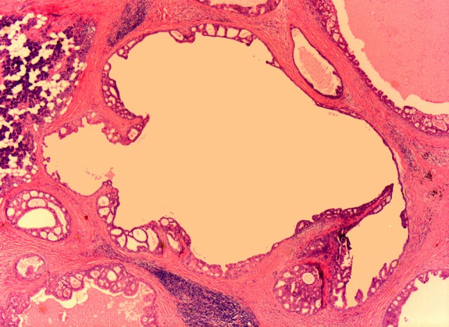
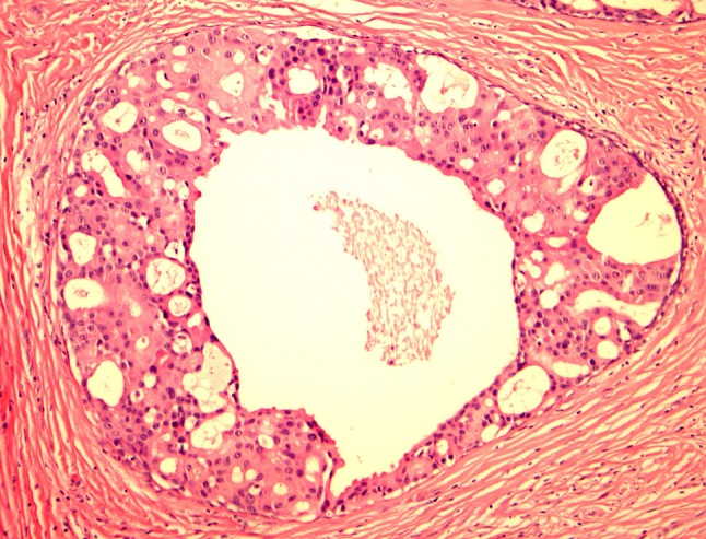
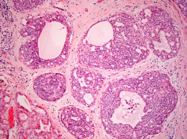
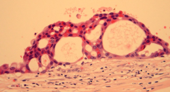
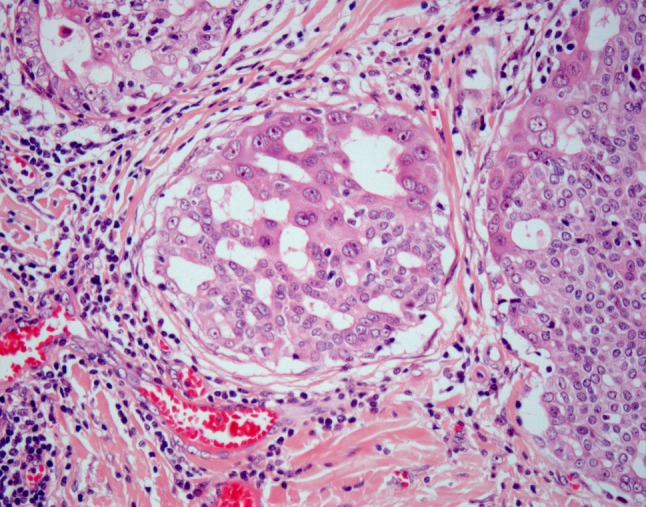
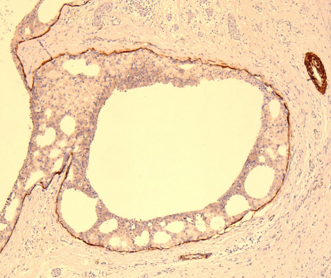
Low-grade salivary duct carcinoma (LG-SDC) is a rare neoplasm characterized by predominant intraductal growth, luminal ductal phenotype, bland microscopic features, and favorable clinical behavior with an appearance reminiscent of florid to atypical ductal hyperplasia to low grade intraductal breast carcinoma. LG-SDC is composed of multiple cysts, cribriform architecture with "Roman Bridges", "pseudocribriform" proliferations with floppy fenestrations or irregular slits, micropapillae with epithelial tufts, fibrovascular cores, and solid areas. Most of the tumor cells are small to medium sized with pale eosinophilic cytoplasm, and round to oval nuclei, which may contain finely dispersed or dark condensed chromatin. Foci of intermediate to high grade atypia, and invasive carcinoma or micro-invasion have been reported in up to 23 % of cases. The neoplastic cells have a ductal phenotype with coexpression of keratins and S100 protein and are surrounded by a layer of myoepithelial cells in non-invasive cases. The main differential diagnosis of LG-SDC includes cystadenoma, cystadenocarcinoma, sclerosing polycystic adenosis, salivary duct carcinoma in situ/high-grade intraductal carcinoma, and papillary-cystic variant of acinic cell carcinoma. There is no published data supporting the continuous classification of LG-SDC as a variant of cystadenocarcinoma. Given that most LG-SDC are non-invasive neoplasms; the terms "cribriform cystadenocarcinoma" and LG-SDC should be replaced by "low-grade intraductal carcinoma" (LG-IDC) of salivary gland or "low-grade intraductal carcinoma with areas of invasive carcinoma" in those cases with evidence of invasive carcinoma.


Figures

Fig. 1
Fig. 1
Low power appearance of LG-SDC showing cysts of various sizes and diameters with a large central cyst surrounded by smaller ones. Note the cribriform architecture with “Roman bridges” and epithelial tufts lining the cysts
Fig. 2
Fig. 2
LG-SDC with round “rigid” cribriform, oval, and slit-like fenestrated spaces. The neoplastic cells are heterogeneous with elongated cells with small dark nuclei, and larger cells with moderate to abundant eosinophilic cytoplasm with round nuclei with visible nucleoli. Apical cytoplasmic blebs characteristic of apocrine metaplasia are readily seen. No distinctive myoepithelial cells are noted
Fig. 3
Fig. 3
LG-SDC with “pseudocribriform” architecture. The ducts and lobules are occupied by a relatively monotonous ductal forming round spaces along with irregular fenestrated secondary lumina. Apocrine metaplasia can be observed in the lower left corner
Fig. 4
Fig. 4
LG-SDC with anastomosing and filigreed epithelial tufts. The tumor cells and exhibit dark nuclei or finely dispersed chromatin. Without immunohistochemical stains it is difficult to see a distinctive myoepithelial layer
Fig. 5
Fig. 5
LG-SDC with streaming elongated cells with dark nuclei or nuclei with fine chromatin and small nuclei. The peripheral myoepithelial cells are barely visible
Fig. 6
Fig. 6
Cellular heterogeneity and cytologic progression in LG-SDC. Most of the neoplastic ductal cells are small with round nuclei containing fine chromatin; however, larger cells with vesicular chromatin and clearly visible nucleoli are also present
Fig. 7
Fig. 7
Calponin highlighting a thin myoepithelial cell layer at the periphery of ducts and cysts in LG-SDC
Fig. 8
Fig. 8
Diffuse expression of S100 protein in a LG-SDC with “pseudocribriform” architecture

Similar articles

Cited by

References

    1. Delgado R, Klimstra DS, Albores-Saavedra J. Low grade salivary duct carcinoma. A distinctive variant with a low grade histology and a predominant intraductal growth pattern. Cancer. 1996;78:958–967. doi: 10.1002/(SICI)1097-0142(19960901)78:5<958::AID-CNCR4>3.0.CO;2-8. - DOI - PubMed
    1. Brandwein-Gensler M, Gnepp DR. Low-grade cribriform cystadenocarcinoma. In: Barnes L, Eveson JW, Reichart P, et al editors. Pathology and Genetics Head and Neck Tumors. Lyon: IARC Press; 2005; 233.
    1. Chen KT. Cytology of salivary duct carcinoma. Diagn Cytopathol. 2000;22:132–135. doi: 10.1002/(SICI)1097-0339(200002)22:2<132::AID-DC17>3.0.CO;2-T. - DOI - PubMed
    1. Brandwein-Gensler M, Hille J, Wang BY, et al. Low-grade salivary duct carcinoma: description of 16 cases. Am J Surg Pathol. 2004;28:1040–1044. doi: 10.1097/01.pas.0000128662.66321.be. - DOI - PubMed
    1. Kusafuka K, Itoh H, Sugiyama C, et al. Low-grade salivary duct carcinoma of the parotid gland: report of a case with immunohistochemical analysis. Med Mol Morphol. 2010;43:178–184. doi: 10.1007/s00795-009-0479-2. - DOI - PubMed

MeSH terms

Substances

LinkOut - more resources