Multicenter validation of spin-density projection-assisted R2-MRI for the noninvasive measurement of liver iron concentration
- PMID: 23821350
- PMCID: PMC4238736
- DOI: 10.1002/mrm.24854
Multicenter validation of spin-density projection-assisted R2-MRI for the noninvasive measurement of liver iron concentration
Abstract
Purpose: Magnetic resonance imaging (MRI)-based techniques for assessing liver iron concentration (LIC) have been limited by single scanner calibration against biopsy. Here, the calibration of spin-density projection-assisted (SDPA) R2-MRI (FerriScan®) in iron-overloaded β-thalassemia patients treated with the iron chelator, deferasirox, for 12 months is validated.
Methods: SDPA R2-MRI measurements and percutaneous needle liver biopsy samples were obtained from a subgroup of patients (n = 233) from the ESCALATOR trial. Five different makes and models of scanner were used in the study.
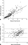
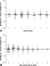
Results: LIC, derived from mean of MRI- and biopsy-derived values, ranged from 0.7 to 50.1 mg Fe/g dry weight. Mean fractional differences between SDPA R2-MRI- and biopsy-measured LIC were not significantly different from zero. They were also not significantly different from zero when categorized for each of the Ishak stages of fibrosis and grades of necroinflammation, for subjects aged 3 to <8 versus ≥8 years, or for each scanner model. Upper and lower 95% limits of agreement between SDPA R2-MRI and biopsy LIC measurements were 74 and -71%.
Conclusion: The calibration curve appears independent of scanner type, patient age, stage of liver fibrosis, grade of necroinflammation, and use of deferasirox chelation therapy, confirming the clinical usefulness of SDPA R2-MRI for monitoring iron overload.
Keywords: ESCALATOR; biopsy; deferasirox; iron overload; β-thalassemia.
Copyright © 2013 Wiley Periodicals, Inc.
Figures


References
-
- Hershko C, Link G, Cabantchik I. Pathophysiology of iron overload. Ann NY Acad Sci. 1998;850:191–201. - PubMed
-
- Brittenham GM, Farrell DE, Harris JW, Feldman ES, Danish EH, Muir WA, Tripp JH, Bellon EM. Magnetic-susceptibility measurement of human iron stores. N Engl J Med. 1982;307:1671–1675. - PubMed
-
- Brittenham GM, Griffith PM, Nienhuis AW, McLaren CE, Young NS, Tucker EE, Allen CJ, Farrell DE, Harris JW. Efficacy of deferoxamine in preventing complications of iron overload in patients with thalassemia major. N Engl J Med. 1994;331:567–573. - PubMed
-
- Brittenham GM, Sheth S, Allen CJ, Farrell DE. Noninvasive methods for quantitative assessment of transfusional iron overload in sickle cell disease. Semin Hematol. 2001;38:37–56. - PubMed
-
- Angelucci E, Baronciani D, Lucarelli G, Baldassarri M, Galimberti M, Giardini C, Martinelli F, Polchi P, Polizzi V, Ripalti M. Needle liver biopsy in thalassaemia: analyses of diagnostic accuracy and safety in 1184 consecutive biopsies. Br J Haematol. 1995;89:757–761. - PubMed
Publication types
MeSH terms
Substances
LinkOut - more resources
Full Text Sources
Other Literature Sources
Medical

