Review
. 2013 Nov;69(2):87-100.
doi: 10.1111/2049-632X.12053. Epub 2013 Jul 2.

Activation of influenza viruses by proteases from host cells and bacteria in the human airway epithelium

Affiliations
Review

Activation of influenza viruses by proteases from host cells and bacteria in the human airway epithelium

Eva Böttcher-Friebertshäuser et al. Pathog Dis. 2013 Nov.

Abstract

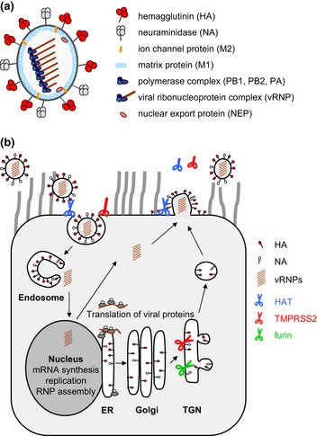
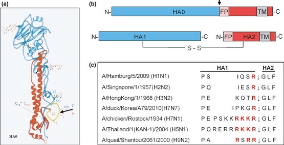
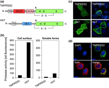
Influenza is an acute infection of the respiratory tract, which affects each year millions of people. Influenza virus infection is initiated by the surface glycoprotein hemagglutinin (HA) through receptor binding and fusion of viral and endosomal membranes. HA is synthesized as a precursor protein and requires cleavage by host cell proteases to gain its fusion capacity. Although cleavage of HA is crucial for virus infectivity, little was known about relevant proteases in the human airways for a long time. Recent progress in the identification and characterization of HA-activating host cell proteases has been considerable however and supports the idea of targeting HA cleavage as a novel approach for influenza treatment. Interestingly, certain bacteria have been demonstrated to support HA activation either by secreting proteases that cleave HA or due to activation of cellular proteases and thereby may contribute to virus spread and enhanced pathogenicity. In this review, we give an overview on activation of influenza viruses by proteases from host cells and bacteria with the main focus on recent progress on HA cleavage by proteases HAT and TMPRSS2 in the human airway epithelium. In addition, we outline investigations of HA-activating proteases as potential drug targets for influenza treatment.

Keywords: HAT; TMPRSS2; influenza treatment by protease inhibitors; influenza virus hemagglutinin; proteolytic cleavage; viral-bacterial pneumonia.


Conflict of interest statement

The authors declare that they have no conflict of interest.

Figures

Figure 1
Figure 1
(a) Scheme of the influenza A virus particle. The virion contains a lipid envelope derived from the cellular plasma membrane. The two glycoproteins hemagglutinin (HA) and neuraminidase (NA) and the ion channel protein M2 are embedded in the envelope. The inner side is lined by the matrix protein M1. The genome consists of eight segments of single‐stranded, negative‐sense RNA, which are associated with the nucleoprotein (NP) and the polymerase subunits PB1, PB2 and PA to form viral ribonucleoprotein complexes (vRNPs). The nuclear export protein (NEP) is also present in the virion, while the nonstructural proteins NS1, PB1‐F2, PB1‐N40, PA‐X, PA‐N155 and PA‐N182 are present in the infected cell. (b) Influenza virus replication and proteolytic activation by cellular proteases in human airway epithelial cells. Infection is initiated by the HA through binding to N‐acetyl neuraminic acid‐containing cell surface receptors. Upon receptor‐mediated endocytosis, HA mediates fusion of viral and endosomal membranes at low pH to release the vRNPs into the cytoplasm (uncoating). The vRNPs are imported into the nucleus, where transcription and replication of the viral genome occur. Translation of viral mRNAs is performed by the cellular machinery. HA, NA and M2 are synthesized into the endoplasmic reticulum (ER) and transported along the constitutive secretory pathway to the plasma membrane. The internal viral proteins are synthesized at free ribosomes, and protein components of the vRNPs are then imported into the nucleus, where assembly of new vRNPs occurs. Finally, vRNPs are exported from the nucleus and transported to the plasma membrane, where self‐assembly of viral proteins leads to budding of new virions. The NA cleaves N‐acetyl neuraminic acid from carbohydrate moieties, facilitating release of progeny virions. Proteolytic activation of HA can take place in different compartments and at different time points during viral replication and is indicated by scissors (open scissor: active protease; closed scissor: enzymatically inactive protease; truncated scissor: soluble protease). HA with multibasic cleavage site is cleaved by furin in the TGN. HA containing a monobasic cleavage site is cleaved by TMPRSS2 in the TGN or by HAT on the plasma membrane: either during assembly and budding of progeny virus or during attachment and entry into the cell.
Figure 2
Figure 2
Cleavage of HA0 into HA1 and HA2 at a specific cleavage site. (a) Structure of the monomeric HA0 precursor of A/HongKong/68 containing the mutation R329Q to prevent cleavage determined by X‐ray crystallography (Chen et al., 1998). The cleavage site is located in a prominent surface loop (yellow) highlighted by an arrow. (b) Schematic illustration of the HA0 precursor and the cleaved form consisting of the disulphide‐linked subunits HA1 and HA2. The colours of HA1 (blue) and HA2 (red) are based on the structure shown in Fig. 1a. The cleavage site is indicated by an arrow. FP: fusion peptide, TM: transmembrane domain. (c) Alignment of amino acid sequences at the HA cleavage site of different human and avian influenza viruses. The arrow indicates the cleavage site between HA1 and HA2. Basic amino acids crucial for cleavage by relevant proteases are highlighted in red.
Figure 3
Figure 3
Human proteases TMPRSS2 and HAT cleave HA with monobasic cleavage site. (a) Schematic domain structures. HAT and TMPRSS2 are synthesized as single‐chain zymogens that consist of an N‐terminal transmembrane domain (TM), a stem region containing, for example, sea urchin sperm protein enterokinase; agrin domain (SEA) for HAT or low‐density lipoprotein receptor class A domain (LDLRA); and scavenger receptor cysteine‐rich domain (SRCR) for TMPRSS2; and a C‐terminal trypsin‐like serine (S1) protease domain with the catalytic triad histidine (H), aspartic acid (D) and serine (S). The zymogens undergo autocatalytic cleavage activation (indicated by arrows), and the catalytic domain remains linked to the transmembrane rest of the molecule by a disulphide bond. (b) Enzymatic activity of cell surface‐anchored or soluble HAT and TMPRSS2 in MDCKHAT and MDCKTMPRSS2 cells. Protease activity on the cell surface and in concentrated protease‐containing supernatants, respectively, was measured by incubation with the fluorogenic peptide substrate Boc‐Gly‐Pro‐Arg‐AMC (Böttcher‐Friebertshäuser et al., 2010). (c) Subcellular localization of HAT and TMPRSS2. HAT and TMPRSS2 on the cell surface (nonpermeabilized cells; left panels) of transient protease expressing MDCK cells and within the cell (permeabilized cells; right panels) were stained using protease‐specific antibodies and FITC‐conjugated secondary antibodies. Cell nuclei were counterstained with DAPI. (d) Colocalization of TMPRSS2 and furin. Huh‐7 cell with transient expression of TMPRSS2 and furin were permeabilized, and protease expression was analysed by TMPRSS2‐ and furin‐specific antibodies, respectively, and FITC‐ or TRITC‐conjugated secondary antibodies. Cell nuclei were counterstained using DAPI.
Figure 4
Figure 4
Activation of influenza viruses by proteases from bacteria. (a) Cleavage of HA of A/swine/1976/31 (H1N1) by proteases from S. aureus. Metabolically S35‐labelled A/swine/1976/31 (H1N1) containing noncleaved HA0 was treated with the indicated proteases or remained untreated (w/o protease). Proteins were separated by SDSPAGE and visualized by autoradiography (Tashiro et al., 1987a). (b) Pathological alterations in the lungs of mice. Mice were infected with either S. aureus Wood 46 or influenza virus A/swine/1976/31 (H1N1) or co‐infected with both pathogens. Noninfected mice were used as a control. Lungs were taken at 5 days p.i. (Tashiro et al., 1987a). Mice indicate survival or fatal infection of the animals. (c) Virus titres in mice lungs. Virus titres in lung homogenates of mice infected with certain influenza viruses and S. aureus strains are shown as plaque‐forming units (p.f.u.) per lung. Lung homogenates were either treated with trypsin or remained untreated before plaque titration. Infections with virus alone without (▵) and with (▲) trypsin treatment; co‐infections without (○) and with (●) trypsin treatment (Tashiro et al., 1987a, 1987b, 1987c). Mice indicate survival and fatal infection, respectively.
Figure 5
Figure 5
Activation of influenza viruses by host cellular and bacterial proteases in the human airway epithelium. Ciliated and nonciliated epithelial cells are shown. HA‐activating proteases are shown by scissors: HAT (blue), TMPRSS2 (red); continuous arrows indicate infection of cells, and dashed arrows, release of virus progeny. (a) Infection of HAT‐ and/or TMPRSS2‐expressing cells results in release of infectious progeny virus containing cleaved HA (green). (b) Infection of cell without expression of relevant HA‐activating proteases results in progeny virus containing noncleaved HA (black). (c) Virus containing noncleaved HA is not able to infect cells expressing only TMPRSS2 or cells without expression of any relevant protease. (d) HAT‐expressing cells support proteolytic activation of HA of incoming virus on the cell surface, facilitating infection of the cell and release of infectious progeny virus. (e) In co‐infections of influenza virus and certain bacteria, soluble bacterial proteases (purple) may support proteolytic activation and spread of the virus. (f) Bacterial proteases or proteins may activate or augment cellular proteases (orange), which in turn cleave HA.

Similar articles

Cited by

References

    1. Afar DE, Vivanco I, Hubert RS, Kuo J, Chen E, Saffran DC, Raitano AB & Jakobovits A (2001) Catalytic cleavage of the androgen‐regulated TMPRSS2 protease results in its secretion by prostate and prostate cancer epithelia. Cancer Res 61: 1686–1692. - PubMed
    1. Baron J, Tarnow C, Mayoli‐Nüssle D et al (2013) Matriptase, HAT, and TMPRSS2 Activate the Hemagglutinin of H9N2 Influenza A Viruses. J Virol 87: 1811–1820. - PMC - PubMed
    1. Becker GL, Sielaff F, Than ME, Lindberg I, Routhier S, Day R, Lu Y, Garten W & Steinmetzer T (2010) Potent inhibitors of furin and furin‐like proprotein convertases containing decarboxylated P1 arginine mimetics. J Med Chem 53: 1067–1075. - PMC - PubMed
    1. Bertram S, Glowacka I, Blazejewska P et al (2010) TMPRSS2 and TMPRSS4 facilitate trypsin‐independent spread of influenza virus in Caco‐2 cells. J Virol 84: 10016–10025. - PMC - PubMed
    1. Bertram S, Heurich A, Lavender H, Gierer S, Danisch S, Perin P, Lucas JM, Nelson PS, Pöhlmann S & Soilleux EJ (2012) Influenza and SARS‐coronavirus activating proteases TMPRSS2 and HAT are expressed at multiple sites in human respiratory and gastrointestinal tracts. PLoS ONE 7: e35876. - PMC - PubMed

Substances