Greatly increased numbers of histamine cells in human narcolepsy with cataplexy
- PMID: 23821583
- PMCID: PMC8211429
- DOI: 10.1002/ana.23968
Greatly increased numbers of histamine cells in human narcolepsy with cataplexy
Abstract
Objective: To determine whether histamine cells are altered in human narcolepsy with cataplexy and in animal models of this disease.
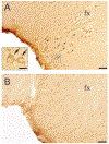
Methods: Immunohistochemistry for histidine decarboxylase (HDC) and quantitative microscopy were used to detect histamine cells in human narcoleptics, hypocretin (Hcrt) receptor-2 mutant dogs, and 3 mouse narcolepsy models: Hcrt (orexin) knockouts, ataxin-3-orexin, and doxycycline-controlled-diphtheria-toxin-A-orexin.
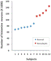
Results: We found an average 64% increase in the number of histamine neurons in human narcolepsy with cataplexy, with no overlap between narcoleptics and controls. However, we did not see altered numbers of HDC cells in any of the animal models of narcolepsy.
Interpretation: Changes in histamine cell numbers are not required for the major symptoms of narcolepsy, because all animal models have these symptoms. The histamine cell changes we saw in humans did not occur in the 4 animal models of Hcrt dysfunction we examined. Therefore, the loss of Hcrt receptor-2, of the Hcrt peptide, or of Hcrt cells is not sufficient to produce these changes. We speculate that the increased histamine cell numbers we see in human narcolepsy may instead be related to the process causing the human disorder. Although research has focused on possible antigens within the Hcrt cells that might trigger their autoimmune destruction, the present findings suggest that the triggering events of human narcolepsy may involve a proliferation of histamine-containing cells. We discuss this and other explanations of the difference between human narcoleptics and animal models of narcolepsy, including therapeutic drug use and species differences.
© 2013 American Neurological Association.
Conflict of interest statement
Potential Conflicts of Interest
Nothing to report.
Figures


References
-
- Peyron C, Faraco J, Rogers W, et al. A mutation in a case of early onset narcolepsy and a generalized absence of hypocretin peptides in human narcoleptic brains. Nat Med 2000;6:991–997. - PubMed
-
- Guilleminault C, Cao MT. Narcolepsy: diagnosis and management. In: Kryger MH, Roth T, Dement WC, eds. Principles and practice of sleep medicine. 5th ed. St Louis, MO: Elsevier Saunders, 2011:957–968.
-
- Lin L, Faraco J, Kadotani H, et al. The sleep disorder canine narcolepsy is caused by a mutation in the hypocretin (orexin) receptor gene. Cell 1999;98:365–376. - PubMed
Publication types
MeSH terms
Substances
Grants and funding
LinkOut - more resources
Full Text Sources
Other Literature Sources

