. 2013 Sep 4;3(9):1453-65.
doi: 10.1534/g3.113.006429.

Mutation rates, spectra, and genome-wide distribution of spontaneous mutations in mismatch repair deficient yeast

Affiliations

Mutation rates, spectra, and genome-wide distribution of spontaneous mutations in mismatch repair deficient yeast

Gregory I Lang et al. G3 (Bethesda). .

Abstract

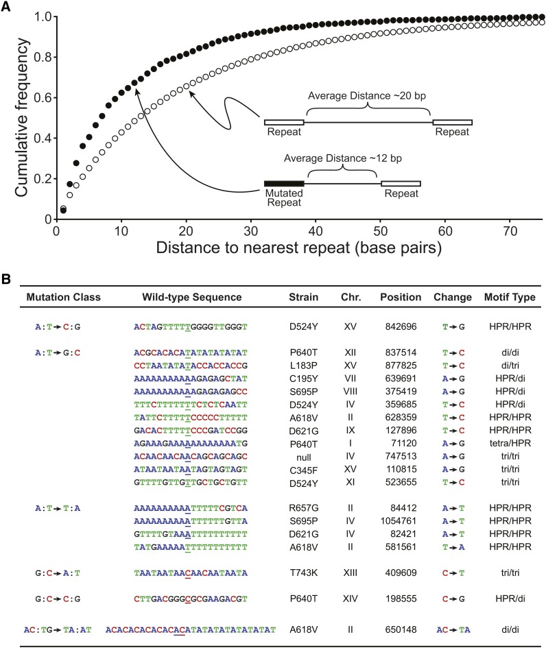
DNA mismatch repair is a highly conserved DNA repair pathway. In humans, germline mutations in hMSH2 or hMLH1, key components of mismatch repair, have been associated with Lynch syndrome, a leading cause of inherited cancer mortality. Current estimates of the mutation rate and the mutational spectra in mismatch repair defective cells are primarily limited to a small number of individual reporter loci. Here we use the yeast Saccharomyces cerevisiae to generate a genome-wide view of the rates, spectra, and distribution of mutation in the absence of mismatch repair. We performed mutation accumulation assays and next generation sequencing on 19 strains, including 16 msh2 missense variants implicated in Lynch cancer syndrome. The mutation rate for DNA mismatch repair null strains was approximately 1 mutation per genome per generation, 225-fold greater than the wild-type rate. The mutations were distributed randomly throughout the genome, independent of replication timing. The mutation spectra included insertions/deletions at homopolymeric runs (87.7%) and at larger microsatellites (5.9%), as well as transitions (4.5%) and transversions (1.9%). Additionally, repeat regions with proximal repeats are more likely to be mutated. A bias toward deletions at homopolymers and insertions at (AT)n microsatellites suggests a different mechanism for mismatch generation at these sites. Interestingly, 5% of the single base pair substitutions might represent double-slippage events that occurred at the junction of immediately adjacent repeats, resulting in a shift in the repeat boundary. These data suggest a closer scrutiny of tumor suppressors with homopolymeric runs with proximal repeats as the potential drivers of oncogenesis in mismatch repair defective cells.

Keywords: homopolymeric runs; microsatellites; mismatch repair; mutation accumulation; mutation rate.


Figures

Figure 1
Figure 1
Mutations in mismatch repair defective cells occur randomly across the genome. (A) Chromosomal distribution of mutations including the single base pair substitutions (open circles) and the insertions/deletion at mono-, di-, and trinucleotide microsatellites (filled circles) are shown at their chromosomal position for each of the 16 yeast chromosomes. Mutation number was plotted against chromosome size for single-base pair substitutions (B) and for insertions/deletions at microsatellites (C). Single-base substitutions in (B) represent data pooled from two independent mutation accumulation experiments. R2 values were generated in Microsoft Excel (Redmond, WA) and are indicated on the graphs.
Figure 2
Figure 2
Mutation rate increases with microsatellite repeat length. The number of insertion/deletion mutations identified at A/T homopolymeric repeats (A), or dinucleotide microsatellites (D) are plotted according to repeat length. Shaded areas indicate that the numbers might be an underrepresentation because of the decreased ability to detect insertions or deletions at long repeats. The number of A/T homopolymers (B) or dinucleotide microsatellites (E) in the yeast genome (y-axis) is plotted according to repeat length (x-axis) on semi-log graphs. The mutation rate (mutation per repeat per generation) for homopolymers (C) or dinucleotide microsatellites (F) are plotted according to repeat unit. The exponential increase in mutation rate from 3 to 8 repeat units is plotted on semi-log graphs in enclosed panels. Formulas and R2 values were generated in Microsoft Excel.
Figure 3
Figure 3
Microsatellites proximal to other repeats are more mutable. (A) The cumulative frequency plots for microsatellites sorted according to the distance to the nearest neighboring repeat for the whole genome (open circles) or for the mutated regions (closed circles) are shown. MATLAB (MathWorks, Inc.) kstest2, Kolmogorov-Smirnov comparison of two data sets, was used to determine the p value, P = 2.8 × 10⁻⁶. The schematic diagram provides an illustration of the relative distance between repeats for the whole genome compared with the mutated microsatellites and the nearest neighboring repeat for a particular point on the graph. (B) The table lists single base substitutions found in regions with immediately adjacent repeats, including homopolymeric runs (HPR), dinucleotide (di), trinucleotide (tri), and tetranucleotide (tetra) microsatellites. The nucleotide sequence is shown and the wild-type base that is mutated in the experimental strain is underlined. The nucleotide change is indicated as is the mutational class. The chromosome position is given for the W303 draft genome (available upon request).
Figure 4
Figure 4
Single-base substitution signature for mismatch repair defective cells. (A) The percentages of each class of single-base substitutions are shown for the pooled mismatch repair defective cells (msh2) and the wild-type reporter construct data (Kunz et al. 1998; Lang and Murray 2008; Ohnishi et al. 2004) compiled by Lynch et al. (i.e., WT Lynch et al.) (Lynch et al. 2008). Transitions and transversions are indicated. The sample size for each strain is given (n). (B) The single-base-pair substitution signatures for the strains completely lacking msh2 function (msh2∆), for the Lynch et al. (2008) wild-type sequencing data (WT seq Lynch et al.) and the wild-type reporter data (WT Lynch et al.) (Kunz et al. 1998; Lang and Murray 2008; Ohnishi et al. 2004) from panel (A) and for strains expressing missense variants of msh2 indicated on the graph as the amino acid substitution (e.g., P640T, proline at codon 640 in the yeast coding sequence is mutated to a threonine). Only signatures that were statistically different (P < 0.01) from the msh2∆ signature using the Fisher exact test (MATLAB script, Guangdi, © 2009) are shown. All but P640L missense substitutions fall in the ATPase domain of Msh2. The sample size for each strain is given (n). Single-base substitutions in this figure represents data pooled from two independent mutation accumulation experiments.

References

    1. Abdulovic A. L., Hile S. E., Kunkel T. A., Eckert K. A., 2011. The in vitro fidelity of yeast DNA polymerase delta and polymerase epsilon holoenzymes during dinucleotide microsatellite DNA synthesis. DNA Repair (Amst.) 10: 497–505 - PMC - PubMed
    1. Acharya S., Wilson T., Gradia S., Kane M. F., Guerrette S., et al. , 1996. hMSH2 forms specific mispair-binding complexes with hMSH3 and hMSH6. Proc. Natl. Acad. Sci. USA 93: 13629–13634 - PMC - PubMed
    1. Agier N., Fischer G., 2012. The mutational profile of the yeast genome is shaped by replication. Mol. Biol. Evol. 29: 905–913 - PubMed
    1. Aquilina G., Bignami M., 2001. Mismatch repair in correction of replication errors and processing of DNA damage. J. Cell. Physiol. 187: 145–154 - PubMed
    1. Arlow T., Scott K., Wagenseller A., Gammie A., 2013. Proteasome inhibition rescues clinically significant unstable variants of the mismatch repair protein Msh2. Proc. Natl. Acad. Sci. USA 110: 246–251 - PMC - PubMed

Publication types

Substances