. 2013 Jul 16;110(29):11863-8.
doi: 10.1073/pnas.1301951110. Epub 2013 Jul 2.

Dynamics of nucleoid structure regulated by mitochondrial fission contributes to cristae reformation and release of cytochrome c

Affiliations

Dynamics of nucleoid structure regulated by mitochondrial fission contributes to cristae reformation and release of cytochrome c

Reiko Ban-Ishihara et al. Proc Natl Acad Sci U S A. .

Abstract

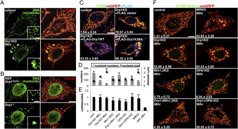
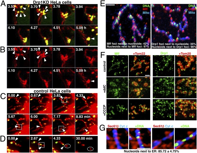
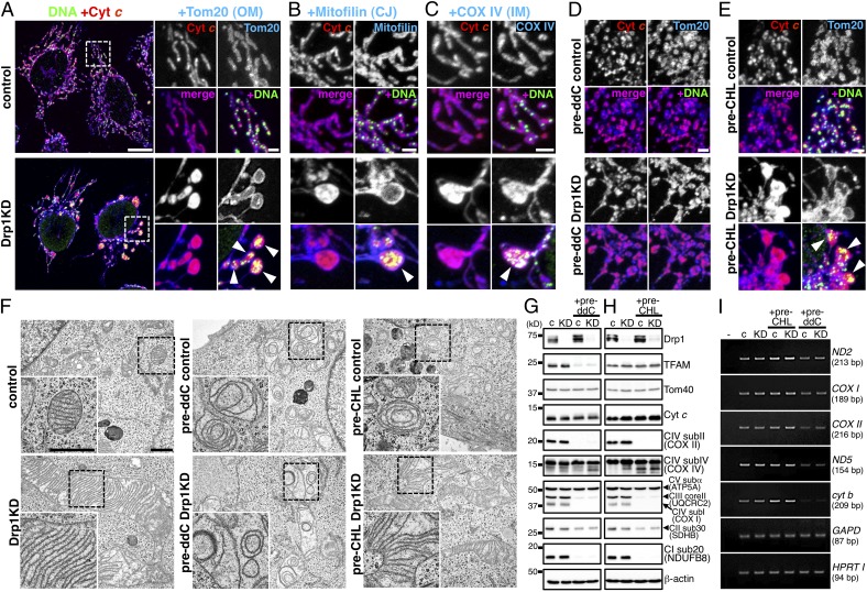
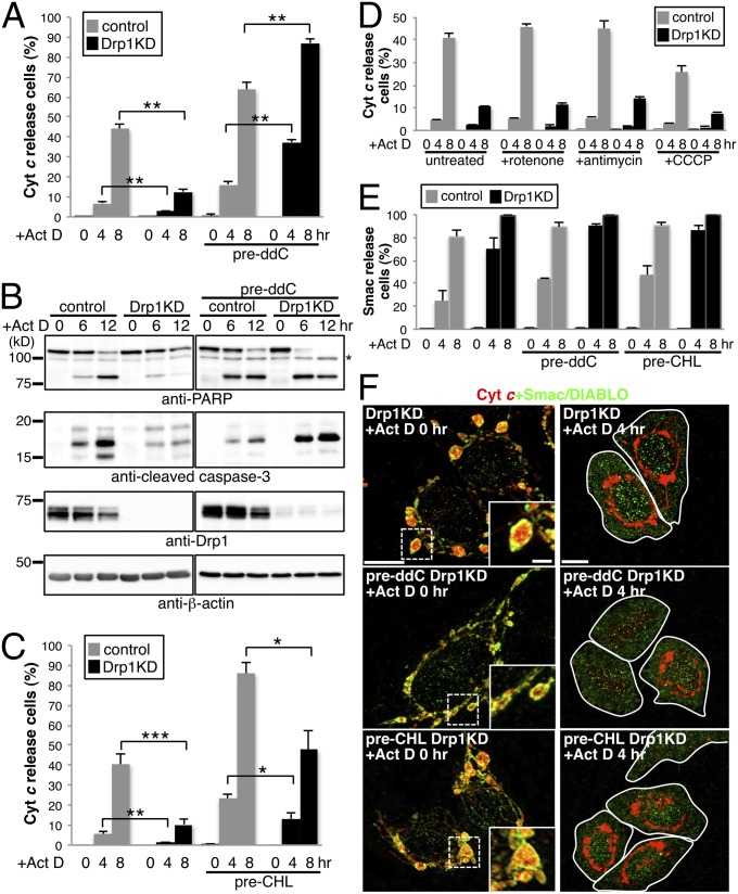
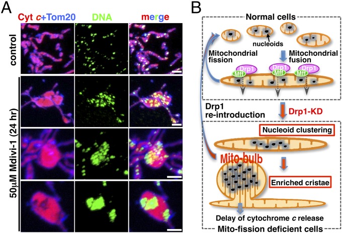
Mammalian cells typically contain thousands of copies of mitochondrial DNA assembled into hundreds of nucleoids. Here we analyzed the dynamic features of nucleoids in terms of mitochondrial membrane dynamics involving balanced fusion and fission. In mitochondrial fission GTPase dynamin-related protein (Drp1)-deficient cells, nucleoids were enlarged by their clustering within hyperfused mitochondria. In normal cells, mitochondrial fission often occurred adjacent to nucleoids, since localization of Mff and Drp1 is dependent on the nucleoids. Thus, mitochondrial fission adjacent to nucleoids should prevent their clustering by maintaining small and fragmented nucleoids. The enhanced clustering of nucleoids resulted in the formation of highly stacked cristae structures in enlarged bulb-like mitochondria (mito-bulbs). Enclosure of proapoptotic factor cytochrome c, but not of Smac/DIABLO, into the highly stacked cristae suppressed its release from mitochondria under apoptotic stimuli. In the absence of nucleoids, Drp1 deficiency failed to form mito-bulbs and to protect against apoptosis. Thus, mitochondrial dynamics by fission and fusion play a critical role in controlling mitochondrial nucleoid structures, contributing to cristae reformation and the proapoptotic status of mitochondria.


Conflict of interest statement

The authors declare no conflict of interest.

Figures

Fig. 1.
Fig. 1.
Enlarged nucleoids in Drp1-deficient cells. (A) Immunofluorescence staining of HeLa cells treated with Drp1 or control siRNA for 96 h as indicated. (B) Drp1 KO and control MEFs. (C) Rescue experiments. KD HeLa cells stably expressing mitRFP were subsequently transfected with WT or K38A of FLAG-tagged rat Drp1 for 20 h. The cells containing enlarged nucleoids were counted in three independent experiments (n = ∼100). (D–F) HeLa cells stably expressing mitRFP were treated with the indicated siRNAs and stained with SYBR Green I. The average of three independent experiments is shown. The average number and diameter of nucleoids in 30 randomly selected cells (D). mtDNA content was quantified by quantitative PCR (E). The cells containing enlarged nucleoids were counted (n = ∼100) (F). Single-color images of A, B, and C are shown in Fig. S1 A and B and Fig. S2A, respectively. (Scale bar: 10 μm; 2 μm in Insets).
Fig. 2.
Fig. 2.
Imaging of mitochondrial fission and nucleoids. (A and B) HeLa cells stably expressing mitRFP treated with Drp1 siRNA for 24 h were stained with SYBR Green I, and live-cell images were obtained by confocal microscopy. The images were obtained every 5 min for 6–10 h as indicated. Magnified images of mitochondria and nucleoids (A) and mitochondria alone (B) in Movie S1 are shown. Red, mitRFP; green, nucleoids (SYBR Green I). Arrowheads indicate clustering of nucleoids. Also see Fig. S3 A–C and Movies S2 and S3. (C and D) Live-cell images of control HeLa cells obtained by CCD camera in Movies S6 (C) and S7 (D). The images were obtained every 10 s. Red, mitRFP; green, nucleoids (SYBR Green I). Arrowheads indicate sites of mitochondrial fission, and dashed squares indicate mitochondrial fission occurring in the vicinity of nucleoids. Dashed circles indicate generated mitochondria devoid of nucleoids. Also see Fig. S3 D–F and Movies S8, S9, and S10. (E) Nucleoids and endogenous Mff or Drp1 localization, shown by intensity profile landscapes. (F) Distribution of Mff and Drp1 in HeLa cells treated with 10 μM ddC for 5 d or with 10 μM CCCP for 45 min. (G) Distribution of ER and nucleoids in control cells. Single-color images of overall landscapes are shown in Fig. S3G. (Scale bar: 2 μm.)
Fig. 3.
Fig. 3.
Mitochondrial cristae were highly stacked in the bulb-like mitochondria (mito-bulbs) with clustered nucleoids. HeLa cells were treated with 10 μM ddC (pre-ddC; D, G, and I) or 100 μM chloramphenicol (pre-CHL; E, H, and I) for 48 h, then further treated with siRNA for Drp1 or control for 96 h in the presence of the same reagents. (A–E) Immunofluorescence staining of nucleoids and mitochondrial proteins: red, cyt c; green, mtDNA; blue, Tom20 (A, D, and E); mitofilin (B); and COX IV (C). Arrowheads indicate mito-bulbs. (Scale bars: 10 μm in A, Left; 2 μm in magnified images in A–E). CJ, cristae junction. Overall landscapes of D and E are shown in Fig. S4 D and E. (F) Images obtained by EM. (Lower, Left) Magnified images of the boxed area in each panel. (Scale bar: 1 μm.) Overall landscapes are shown in Fig. S4 F and G. (G and H) Mitochondrial protein levels determined by immunoblot analysis using the indicated antibodies in ddC-treated (G) and CHL-treated (H) cells. Several subunits of OXPHOS complexes, such as the mitochondria-encoded COX I and II (complex IV), as well as nuclear genome-encoded respiratory proteins, such as NDUFB8 (complex I) and UQCRC2 (complex III), were repressed. Nuclear genome-encoded SDHB (complex II), COX IV (complex IV), and ATP5A (complex V) were less affected. (I) RNAs by RT-PCR. Mitochondrial transcripts (ND2, ND5, COX I, COX II, and cyt b) and nuclear genome-encoded transcripts (GAPD and HPRT I) were analyzed. c, control; KD, Drp1 KD.
Fig. 4.
Fig. 4.
Nucleoid clustering was required for the apoptosis delay in Drp1-deficient cells. HeLa cells treated as described in Fig. 3 were further treated with 5 μM Act D, with (A, C, D, and E) or without (B) 50 μM zVAD-fmk for the indicated periods. (A, C, and D) Cyt c and Tom20 were stained, and cells with released cyt c were counted. (E and F) Smac/DIABLO and cyt c were stained, and cells with released Smac/DIABLO were counted. At least 300 cells in three independent experiments were counted. Data are expressed as mean ± SD. *P < 0.05; **P < 0.01; ***P < 0.001. (B) PARP and cleaved caspase-3 were measured by immunoblot analysis. The asterisk indicates a nonspecific band. (D) Effect of respiratory inhibitors on apoptosis. Drp1 KD cells were treated with 25 μM rotenone (complex I inhibitor), 1 μM antimycin (complex III inhibitor), and 10 μM CCCP (uncoupler) for 1 h. (E and F) Cyt c (red) and Smac/DIABLO (green) were immunostained (F), and the cells with released Smac/DIABLO were counted (E). (Scale bars: 10 μm; 2 μm in Insets).
Fig. 5.
Fig. 5.
Mitochondrial fission regulates nucleoid clustering, mito-bulb formation with IM cristae, and apoptosis. (A) Nucleoids (anti-DNA, green), cyt c (red), and Tom20 (blue) were immunostained in HeLa cells treated with or without Drp1 inhibitor Mdivi-1 for 24 h. (Scale bar: 2 μm.) (B) Schematic representation of our results (Discussion).

References

    1. Lang BF, et al. An ancestral mitochondrial DNA resembling a eubacterial genome in miniature. Nature. 1997;387(6632):493–497. - PubMed
    1. Kuroiwa T. Mechanisms of organelle division and inheritance and their implications regarding the origin of eukaryotic cells. Proc Jpn Acad, Ser B, Phys Biol Sci. 2010;86(5):455–471. - PMC - PubMed
    1. Garrido N, et al. Composition and dynamics of human mitochondrial nucleoids. Mol Biol Cell. 2003;14(4):1583–1596. - PMC - PubMed
    1. Legros F, Malka F, Frachon P, Lombès A, Rojo M. Organization and dynamics of human mitochondrial DNA. J Cell Sci. 2004;117(Pt 13):2653–2662. - PubMed
    1. Iborra FJ, Kimura H, Cook PR. The functional organization of mitochondrial genomes in human cells. BMC Biol. 2004;2:9. - PMC - PubMed

Publication types

MeSH terms